
Page 1086 - Small Animal Internal Medicine, 6th Edition
P. 1086
1058 PART IX Nervous System and Neuromuscular Disorders
Medial rectus Dorsal oblique (CN4)
Dorsal oblique (CN3) Dorsal rectus (CN3)
VetBooks.ir Dorsal rectus
Lateral rectus Lateral rectus (CN4)
Ventral rectus Ventral rectus (CN3)
Ventral oblique Ventral oblique (CN3)
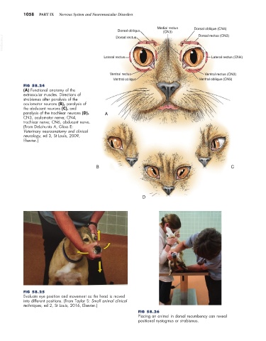
FIG 58.24
(A) Functional anatomy of the
extraocular muscles. Directions of
strabismus after paralysis of the
oculomotor neurons (B), paralysis of
the abducent neurons (C), and
paralysis of the trochlear neurons (D). A
CN3, oculomotor nerve; CN4,
trochlear nerve; CN6, abducent nerve.
(From DeLahunta A, Glass E:
Veterinary neuroanatomy and clinical
neurology, ed 3, St Louis, 2009,
Elsevier.)
B C
D
FIG 58.25
Evaluate eye position and movement as the head is moved
into different positions. (From Taylor S: Small animal clinical
techniques, ed 2, St Louis, 2016, Elsevier.)
FIG 58.26
Placing an animal in dorsal recumbency can reveal
positional nystagmus or strabismus.