
Page 1089 - Small Animal Internal Medicine, 6th Edition
P. 1089
CHAPTER 58 Lesion Localization and the Neurologic Examination 1061
LESION LOCALIZATION hunting, herding, racing, jumping) may be at increased risk
After the neurologic examination is completed, an animal’s for specific activity-related injuries. Potential exposure to
VetBooks.ir mentation, cranial nerves, posture, gait, forelimbs, rear trauma, toxins, and infectious disorders should be ascer-
tained through careful history taking.
limbs, perineum, anus, and bladder can be characterized as
normal or abnormal. If disease above the foramen magnum
is present, clinical findings should allow a lesion to be local- DISEASE ONSET AND PROGRESSION
ized to a specific region of the brain. In patients with spinal Evaluation of the onset and progression of neurologic signs
cord disease, determining whether the neurologic abnormal- is of primary importance in prioritizing the list of differential
ity in each limb is UMN or LMN in origin allows localiza- diagnoses (Box 58.10). The signs may be peracute and non-
tion to a region of the spinal cord or specific spinal cord progressive, or they may become progressively more severe
segments (see Box 58.4). When LMN signs are present in with time. In peracute disorders, the time of onset of the
a single limb, the lesion can often be even more precisely neurologic signs can be pinpointed exactly, with the animal
localized by determining the muscles affected and, if sensory going from being normal to abnormal within minutes or
nerves are also affected, by testing sensation in dermatomes. hours. Signs reach maximal intensity very rapidly and then
Focal hyperpathia may also help to precisely localize a lesion. remain static or improve over time. Examples include exter-
Whenever possible, the clinician should be able to explain nal trauma, internal trauma from intervertebral disk extru-
all detected neurologic abnormalities on the basis of a single sion, vascular disorders such as infarcts or hemorrhage, and
lesion. Occasionally, however, this will be impossible because some rapid-acting intoxications such as strychnine. Rarely,
the animal has multiple foci of disease or a diffuse disorder. animals with a typically slow progressive disorder (e.g.,
tumor) present with a peracute exacerbation of their signs as
a result of hemorrhage or fracture at the site of the tumor. A
DIAGNOSTIC APPROACH thorough history will often reveal that these animals were
not entirely normal before the acute deterioration.
Once a neurologic lesion has been localized, it is necessary Neurologic disorders with fairly rapid deterioration over
to generate a list of likely differential diagnoses. This list days to weeks are classified as subacute and progressive.
should take into account the signalment, historical data, the Infectious and noninfectious inflammatory diseases and
neuroanatomic location of the lesion, and the nature of the some of the more rapidly progressive neoplasms (e.g., lym-
onset and progression of neurologic signs. It is important to phomas, metastatic malignancies) usually fall into this
consider all possible mechanisms or causes of disease that category. Metabolic and nutritional disorders and some
can affect the nervous system (Box 58.9). Once a list of likely intoxications can also cause subacute progressive signs.
differential diagnoses has been developed, diagnostic tests Animals with chronic progressive signs that develop very
can be performed to confirm or exclude each. slowly over many weeks or months are most likely to have
neoplastic or degenerative disease.
ANIMAL HISTORY
Patient age, gender, breed, and lifestyle may provide clues
regarding the underlying disease. Young animals are most
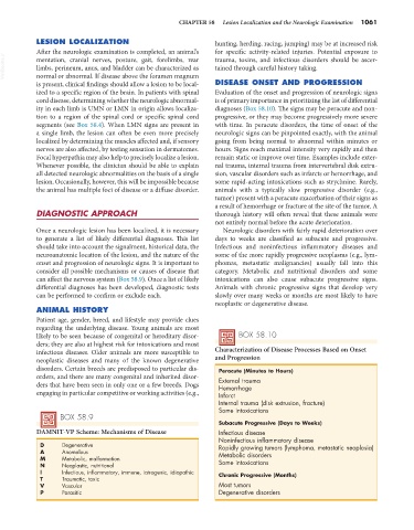
likely to be seen because of congenital or hereditary disor- BOX 58.10
ders; they are also at highest risk for intoxications and most
infectious diseases. Older animals are more susceptible to Characterization of Disease Processes Based on Onset
neoplastic diseases and many of the known degenerative and Progression
disorders. Certain breeds are predisposed to particular dis- Peracute (Minutes to Hours)
orders, and there are many congenital and inherited disor- External trauma
ders that have been seen in only one or a few breeds. Dogs Hemorrhage
engaging in particular competitive or working activities (e.g., Infarct
Internal trauma (disk extrusion, fracture)
Some intoxications
BOX 58.9
Subacute Progressive (Days to Weeks)
DAMNIT-VP Scheme: Mechanisms of Disease Infectious disease
Noninfectious inflammatory disease
D Degenerative Rapidly growing tumors (lymphoma, metastatic neoplasia)
A Anomalous Metabolic disorders
M Metabolic, malformation Some intoxications
N Neoplastic, nutritional
I Infectious, inflammatory, immune, iatrogenic, idiopathic Chronic Progressive (Months)
T Traumatic, toxic
V Vascular Most tumors
P Parasitic Degenerative disorders