
Page 1094 - Small Animal Internal Medicine, 6th Edition
P. 1094
1066 PART IX Nervous System and Neuromuscular Disorders
VetBooks.ir
L1
L2
L1
L2
A B
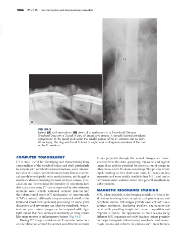
FIG 59.2
Lateral (A) and ventrodorsal (B) views of a myelogram in a 5-month-old German
Shepherd Dog with a 3-week history of progressive ataxia. A dorsally located extradural
compression of the spinal cord within the caudal portion of the L1 vertebra can be seen.
At necropsy, the dog was found to have a single focal cartilaginous exostosis of the roof
of the L1 vertebra.
COMPUTED TOMOGRAPHY X-rays projected through the animal. Images are recon-
CT is most useful for identifying and characterizing bony structed from this data, generating transverse and sagittal
abnormalities of the vertebral bodies and skull, particularly image slices and the potential for construction of images in
in patients with vertebral fractures/luxations, acute mineral- other planes and 3-D volume renderings. This process is very
ized disk extrusions, vertebral tumors, bony lesions of cervi- rapid, resulting in very short scan times. CT scans are less
cal spondylomyelopathy, otitis media/interna, and fungal or expensive and more readily available than MRI, and can be
neoplastic disease involving the nasal cavity or sinuses. Visu- performed under sedation rather than general anesthesia in
alization and determining the laterality of nonmineralized stable patients.
disk extrusions using CT can be improved by administering
nonionic water soluble iodinated contrast material into MAGNETIC RESONANCE IMAGING
the subarachnoid space (CT-myelogram) or intravenously MRI, when available, is the imaging modality of choice for
(CT-IV contrast). Although intraparenchymal detail of the all lesions involving brain or spinal cord parenchyma, and
brain and spinal cord is generally poor using CT alone, gross peripheral nerves. MR images provide excellent soft tissue
distortions and asymmetry can often be visualized. Precon- contrast resolution, displaying excellent neuroanatomical
trast and postcontrast images can be compared with high- detail while providing insight into tissue composition and
light lesions that have increased vascularity or leaky vessels response to injury. The appearance of brain lesions using
like many tumors or inflammatory lesions (Fig. 59.3) different MRI sequences not only localizes lesions precisely
During CT image acquisition an X-ray tube moves in a but helps distinguish inflammatory, neoplastic, and hemor-
circular direction around the animal, and detectors measure rhagic lesions and infarcts. In animals with brain tumors,