
Page 1099 - Small Animal Internal Medicine, 6th Edition
P. 1099
CHAPTER 59 Diagnostic Tests for Nervous System and Neuromuscular Disorders 1071
Most of the cells in the CSF of normal dogs and cats are
small, well-differentiated lymphocytes (60%-70%). Mini-
VetBooks.ir mally vacuolated large mononuclear phagocytes comprise
most of the additional cells. Occasional neutrophils and
S eosinophils are present, but these cells should not normally
make up more than 2% of the cell population. The typical
L1 L2 L3 L4 L5 L6 L7
CSF findings in some specific disorders in dogs and cats are
summarized in Box 59.3. It is important to realize, however,
that CSF cytologic findings must always be interpreted in
relation to the signalment, history, and clinical findings.
If blood contamination is severe, it can influence the cyto-
logic findings, but even grossly apparent iatrogenic contami-
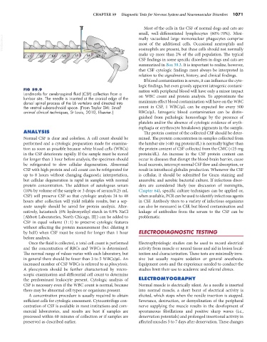
FIG 59.9 nation with peripheral blood will have only a minor impact
Landmarks for cerebrospinal fluid (CSF) collection from a
lumbar site. The needle is inserted at the cranial edge of the on WBC count and protein analysis. To approximate the
dorsal spinal process of the L6 vertebra and directed into maximum effect blood contamination will have on the WBC
the ventral subarachnoid space. (From Taylor SM: Small count in CSF, 1 WBC/µL can be expected for every 500
animal clinical techniques, St Louis, 2010, Elsevier.) RBCs/µL. Iatrogenic blood contamination can be distin-
guished from pathologic hemorrhage by the presence of
platelets and/or the absence of cytologic evidence of eryth-
rophagia or erythrocyte breakdown pigments in the sample.
ANALYSIS The protein content of the collected CSF should be deter-
Normal CSF is clear and colorless. A cell count should be mined. The protein concentration in samples collected from
performed and a cytologic preparation made for examina- the lumbar site (<40 mg protein/dL) is normally higher than
tion as soon as possible because white blood cells (WBCs) the protein content of CSF collected from the CMC (<25 mg
in the CSF deteriorate rapidly. If the sample must be stored protein/dL). An increase in the CSF protein content can
for longer than 1 hour before analysis, the specimen should occur in diseases that disrupt the blood-brain barrier, cause
be refrigerated to slow cellular degeneration. Abnormal local necrosis, interrupt normal CSF flow and absorption, or
CSF with high protein and cell count can be refrigerated for result in intrathecal globulin production. Whenever the CSF
up to 8 hours without changing diagnostic interpretation, is cellular, it should be submitted for Gram staining and
but cellular degeneration is rapid in samples with normal anaerobic and aerobic bacterial culture. If infectious disor-
protein concentration. The addition of autologous serum ders are considered likely (see discussion of meningitis,
(10% by volume of the sample or 3 drops of serum/0.25 mL Chapter 64), specific culture techniques can be applied or,
CSF) will preserve CSF so that cytologic analysis 24 to 48 when available, PCR can be used to identify infectious agents
hours after collection will yield reliable results, but a sep- in CSF. Antibody titers to a variety of infectious organisms
arate sample should be saved for protein analysis. Alter- can also be measured in CSF, but blood contamination and
natively, hetastarch (6% hydroxyethyl starch in 0.9% NaCl leakage of antibodies from the serum to the CSF can be
[Abbott Laboratories, North Chicago, Ill]) can be added to problematic.
CSF in equal volume (1 : 1) to preserve cytologic features
without affecting the protein measurement (but diluting it
by half) when CSF must be stored for longer than 1 hour ELECTRODIAGNOSTIC TESTING
before analysis.
Once the fluid is collected, a total cell count is performed Electrophysiologic studies can be used to record electrical
and the concentration of RBCs and WBCs is determined. activity from muscle or neural tissue and aid in lesion local-
The normal range of values varies with each laboratory, but ization and characterization. These tests are minimally inva-
in general there should be fewer than 3 to 5 WBCs/µL. An sive but usually require sedation or general anesthesia.
increased number of CSF WBCs is referred to as pleocytosis. Equipment costs and the experience needed to conduct the
A pleocytosis should be further characterized by micro- studies limit their use to academic and referral clinics.
scopic examination and differential cell count to determine
the predominant leukocyte present. Cytologic analysis of ELECTROMYOGRAPHY
CSF is necessary even if the WBC count is normal, because Normal muscle is electrically silent. As a needle is inserted
there may be abnormal cell types or organisms present. into normal muscle, a short burst of electrical activity is
A concentration procedure is usually required to obtain elicited, which stops when the needle insertion is stopped.
sufficient cells for cytologic assessment. Cytocentrifuge con- Severance, destruction, or demyelination of the peripheral
centration of CSF is available in most institutions and com- nerve supplying the muscle results in the development of
mercial laboratories, and results are best if samples are spontaneous fibrillations and positive sharp waves (i.e.,
processed within 60 minutes of collection or if samples are denervation potentials) and prolonged insertional activity in
preserved as described earlier. affected muscles 5 to 7 days after denervation. These changes