Page 1103 - Small Animal Internal Medicine, 6th Edition
P. 1103
CHAPTER 60   Intracranial Disorders   1075


            normal cerebellar regulation of the rate, range, and force of   complete physical and neurologic examination as well as an
            movement. Animals with disease limited to the cerebellum   ophthalmologic examination. When the cause of the neuro-
  VetBooks.ir  are ataxic but strong, with normal postural reactions and   logic signs is not readily apparent, animals should be screened
                                                                 for metabolic disorders and systemic manifestations of infec-
            spinal reflexes. They are unable to judge distances or control
            the range of movements and will make a series of jerking and
                                                                 cytologic examination of aspirates from lymph nodes and
            bobbing movements (intention tremor) when attempting to   tious or neoplastic disease using clinicopathologic tests,
            perform precise movements. A fine tremor of the head and   masses, thoracic and abdominal radiographs, and abdominal
            body may also be present at rest.                    ultrasound. Approximately 10% of dogs with intracranial
                                                                 neoplasia have abnormalities on these staging tests that make
            DIAGNOSTIC APPROACH TO ANIMALS                       further diagnostic evaluation unnecessary. Advanced neuro-
            WITH INTRACRANIAL DISEASE                            imaging (computed tomography [CT] or magnetic reso-
                                                                 nance imaging [MRI]) and cerebrospinal fluid (CSF)
            Intracranial disorders that commonly cause neurologic signs   collection and analysis will be required for diagnosis when
            include external trauma, vascular disorders (e.g., hemor-  disease is restricted to an intracranial site. MRI provides
            rhage and infarction), congenital anomalies (e.g., hydro-  much better soft tissue resolution than CT and is the pre-
            cephalus, lissencephaly, cerebellar hypoplasia), infectious   ferred imaging method whenever brain lesions are suspected,
            and noninfectious inflammatory diseases (e.g., encephalitis),   but CT is faster, less expensive, and more readily available in
            degenerative disorders, and primary or metastatic brain   emergency situations, and it provides adequate images when
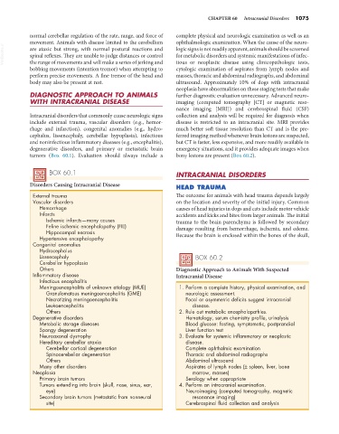
            tumors (Box 60.1). Evaluation should always include a   bony lesions are present (Box 60.2).


                   BOX 60.1                                      INTRACRANIAL DISORDERS

            Disorders Causing Intracranial Disease
                                                                 HEAD TRAUMA
             External trauma                                     The outcome for animals with head trauma depends largely
             Vascular disorders                                  on the location and severity of the initial injury. Common
               Hemorrhage                                        causes of head injuries in dogs and cats include motor vehicle
               Infarcts                                          accidents and kicks and bites from larger animals. The initial
                  Ischemic infarcts—many causes                  trauma to the brain parenchyma is followed by secondary
                  Feline ischemic encephalopathy (FIE)           damage resulting from hemorrhage, ischemia, and edema.
                  Hippocampal necrosis
               Hypertensive encephalopathy                       Because the brain is enclosed within the bones of the skull,
             Congenital anomalies
               Hydrocephalus
               Lissencephaly                                            BOX 60.2
               Cerebellar hypoplasia
               Others                                            Diagnostic Approach to Animals With Suspected
             Inflammatory disease                                Intracranial Disease
               Infectious encephalitis
               Meningoencephalitis of unknown etiology (MUE)      1. Perform a complete history, physical examination, and
                  Granulomatous meningoencephalitis (GME)           neurologic assessment.
                  Necrotizing meningoencephalitis                   Focal or asymmetric deficits suggest intracranial
                  Leukoencephalitis                                   disease.
                  Others                                          2. Rule out metabolic encephalopathies.
             Degenerative disorders                                 Hematology, serum chemistry profile, urinalysis
               Metabolic storage diseases                           Blood glucose: fasting, symptomatic, postprandial
               Spongy degeneration                                  Liver function test
               Neuroaxonal dystrophy                              3. Evaluate for systemic inflammatory or neoplastic
               Hereditary cerebellar ataxia                         disease.
                  Cerebellar cortical degeneration                  Complete ophthalmic examination
                  Spinocerebellar degeneration                      Thoracic and abdominal radiographs
                  Others                                            Abdominal ultrasound
               Many other disorders                                 Aspirates of lymph nodes (± spleen, liver, bone
             Neoplasia                                                marrow, masses)
               Primary brain tumors                                 Serology when appropriate
               Tumors extending into brain (skull, nose, sinus, ear,   4. Perform an intracranial examination.
                  eye)                                              Neuroimaging (computed tomography, magnetic
               Secondary brain tumors (metastatic from nonneural      resonance imaging)
                  site)                                             Cerebrospinal fluid collection and analysis

