
Page 1107 - Small Animal Internal Medicine, 6th Edition
P. 1107
CHAPTER 60 Intracranial Disorders 1079
or fontanelles that remain open at 9 weeks of age do have or closed, ultrasound scanning is more difficult but may still
some ventricular dilation, many will never develop clinical be attempted through the temporal bone in young animals.
VetBooks.ir signs of hydrocephalus. Alternatively, CT or MRI can be performed to detect ven-
Animals with symptomatic hydrocephalus can be slow
tricular enlargement. Although historical studies have shown
learners and may be difficult to housetrain. They may seem
signs, one report showed that ventricular enlargement
dull or depressed. They may have episodic or constant abnor- very little correlation between ventricular size and clinical
mal behavior, circling, and cortical blindness. Seizures may (ventricle/brain [VB] ratio) was correlated with severity of
occur. Severely affected animals may exhibit tetraparesis, clinical signs in small-breed dogs and that all asymptomatic
slow postural reactions, head tilt, or nystagmus. Some puppies with a VB ratio of over 60% went on to develop
animals will develop a ventrolateral strabismus (see Fig. neurologic signs related to their hydrocephalus.
60.1) due either to malformation of the orbit or brainstem Long-term medical management of animals with neuro-
dysfunction. Neurologic signs have an unpredictable course; logic signs is directed at limiting CSF production and reduc-
deficits can progress over time, remain static, and even ing intracranial pressure. Acetazolamide (10 mg/kg orally
improve after 1 to 2 years of age. Signs can worsen coincident q8h), alone or in combination with oral furosemide (1 mg/
with other diseases or minor head trauma. About 30% of kg/day) is the most commonly used drug therapy. Omepra-
dogs with congenital hydrocephalus are not overtly symp- zole may also decrease CSF production and aid control. Some
tomatic until 2 years of age. animals improve with glucocorticoid treatment (prednisone,
Hydrocephalus is suspected on the basis of characteristic 0.5 mg/kg, administered orally daily, tapered weekly until
signs and physical examination findings in a young animal 0.1 mg/kg q48h). Seizures may be controlled with antiepilep-
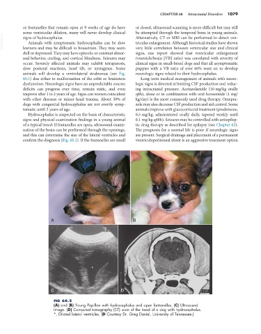
of a typical breed. If fontanelles are open, ultrasound exami- tic drug therapy as described for epilepsy (see Chapter 62).
nation of the brain can be performed through the openings, The prognosis for a normal life is poor if neurologic signs
and this can determine the size of the lateral ventricles and are present. Surgical drainage and placement of a permanent
confirm the diagnosis (Fig. 60.2). If the fontanelles are small ventriculoperitoneal shunt is an aggressive treatment option
A B
*
*
C D
FIG 60.2
(A) and (B) Young Papillon with hydrocephalus and open fontanelles. (C) Ultrasound
image. (D) Computed tomography (CT) scan of the head of a dog with hydrocephalus.
*, Dilated lateral ventricles. (D Courtesy Dr. Greg Daniel, University of Tennessee.)