
Page 1104 - Small Animal Internal Medicine, 6th Edition
P. 1104
1076 PART IX Nervous System and Neuromuscular Disorders
as brain volume increases with edema or hemorrhage, there (SoluMedrol) during the first 6 hours after presentation has
is an increase in intracranial pressure, leading to decreased been shown to be beneficial in patients with spinal cord
VetBooks.ir cerebral perfusion and further brain damage. injury but may actually be detrimental in patients with
serious brain injury.
Initial management of a patient with brain injury should
Systemic and neurologic assessment should be repeated
focus on recognizing and treating systemic injuries and
maintaining adequate circulation and respiration. Systemic every 30 minutes. Although a scoring system (modified
hypotension decreases cerebral perfusion, so fluids should Glasgow Coma Scale) has been developed to allow grading
be administered to maintain blood volume (Box 60.3). and serial monitoring of neurologic status, it is only useful
Administration of synthetic colloids such as Pentaspan (10% in dogs with such severe brainstem and cerebral injury that
Pentastarch in 0.9% NaCL [Bristol-Myers Squibb]) or they are unlikely to recover.
administration of hypertonic saline solution (7.2%) allows
rapid restoration of blood volume and pressure without the VASCULAR ACCIDENTS
large volume of fluid required when crystalloids alone are Vascular accidents, also called strokes, occasionally occur in
administered. Oxygen should be administered by mask or the central nervous systems (CNS) of dogs and cats, and
via nasal or transtracheal catheter. If the animal is uncon- cause a peracute onset of neurologic abnormalities. Ischemic
scious, immediate intubation and ventilation may be strokes result from occlusion of an intracranial blood vessel
required. Hyperventilation reduces intracranial pressure but by a thrombus or embolus. Hemorrhagic strokes result from
causes cerebral vasoconstriction and decreased cerebral per- bleeding into the brain parenchyma.
fusion, so it must be used with caution. Whenever possible, Ischemic strokes occur when cells or clots enter the cir-
a PaCO 2 of 30 to 35 mm Hg should be maintained. If seizures culation from septic foci (endocarditis), primary or meta-
are evident, aggressive anticonvulsant therapy should be ini- static neoplasia, vasculitis, heartworm disease, or cardiac
tiated as for status epilepticus (see Chapter 62) because disease. Atherosclerosis, a common cause of thrombo-
seizure activity greatly increases intracranial pressure. Mea- embolic stroke in humans, has also been associated with
sures to lower intracranial pressure include elevating the stroke due to intravascular clot formation in dogs with
head at a 30-degree angle from horizontal, administration of hypothyroidism, and less commonly in dogs with diabe-
an osmotic diuretic such as intravenous mannitol (1 to 1.5 g/ tes mellitus, hyperadrenocorticism, or hereditary hyper-
kg over 15 minutes) or hypertonic saline solution (4-5 mL/ lipidemia. Chronic hypertension also promotes vascular
kg of 7.2% NaCl over 2-5 minutes), and administration of changes predisposing to ischemic or hemorrhagic stroke.
narcotic analgesic medications as required. Administration Animals with peracute neurologic signs should always have
of high doses of methylprednisolone sodium succinate their blood pressure measured, and if they are hypertensive
they should be evaluated for potential causes such as renal
failure, hyperadrenocorticism (dogs), and hyperthyroid-
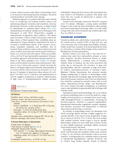
BOX 60.3 ism (cats). Despite thorough investigations, an underlying
cause is only identified in approximately half of all dogs with
Management of Intracranial Injury ischemic stroke.
Spontaneous intracranial hemorrhage can occur without
All Patients an obvious underlying cause or can be the result of a coagu-
Establish patent airway, administer oxygen. lopathy, a bleeding primary or metastatic brain tumor (espe-
Examine, assess, and treat concurrent injuries. cially hemangiosarcoma), or hypertension. Screening for
Treat shock, maintain blood pressure (intravenous fluids, thrombocytopenia, inherited or acquired coagulopathies,
colloids). disseminated intravascular coagulation, systemic neoplasia,
Isotonic crystalloids: 1 4 of shock dose (90 mL/kg dog, and disorders associated with hypertension is indicated
60 mL/kg cat), reassess q15min
Pentaspan: 1 4 of the 20 mL/kg shock dose when intracranial bleeding is suspected.
Hypertonic saline: 4-5 mL/kg of 7.2% NaCl over 2-5 In ischemic or hemorrhagic stroke, the abruptness with
minutes which neurologic deficits develop is highly suggestive of a
Maintain mean arterial blood pressure 80-120 mm Hg. vascular disorder. There may be some progression of signs
Monitor neurologic status every 30 minutes. due to edema in the first 24 to 72 hours, but most animals
with nonfatal ischemic strokes begin to recover quickly.
If Severe Initial Injury or Deterioration Hemorrhagic strokes are more likely than ischemic strokes
Elevate head 30 degrees. to cause rapidly progressive neurologic deficits, increased
Treat seizures if present (see Box 62.7). intracranial pressure, and death.
Administer hyperosmolar therapy. Neurologic deficits reflect the location of the vascular
20% mannitol: 1-1.5 g/kg, administered intravenously insult, with forebrain and cerebellar strokes most common
over 15 minutes (can repeat in 3 hours) or
Hypertonic saline: 4-5 mL/kg of 7.2% NaCl over 2-5 in dogs. In cats, forebrain and brainstem infarcts are more
minutes common. A predisposition for cerebellar infarcts is present
If intubated, maintain PaCO 2 at 30-35 mm Hg. in hypothyroid hyperlipidemic Labrador Retrievers and
in Cavalier King Charles Spaniels. Results of physical