
Page 1106 - Small Animal Internal Medicine, 6th Edition
P. 1106
1078 PART IX Nervous System and Neuromuscular Disorders
Early reports suggested that FHN had a poor prognosis,
as all cats died or were euthanized when initial anticon-
VetBooks.ir vulsant therapy was ineffective, neurologic and behavioral
abnormalities persisted, or recurrent seizures were difficult
to control. There have, however, been reports of a few cats
with FHN that gradually returned nearly to normal with few
neurologic deficits and well-controlled seizures, suggesting
that the long-term outcome can occasionally be good to
excellent. There is some speculation that FHN could be a
manifestation of autoimmune limbic encephalitis following
reports that many cats with FHN experienced full remission
(seizure-free) after antiepileptic, supportive, and corticoste-
roid treatment.
THIAMINE DEFICIENCY
Thiamine (vitamin B1) is a water-soluble vitamin that is A
important in normal carbohydrate metabolism and synthe-
sis of neurotransmitters. Cats require more thiamine than
dogs and can develop thiamine deficiency due to prolonged
anorexia, maldigestion /malabsorption disorders, inadequate
dietary intake, or ingestion of thiaminase in raw fish. Most
cases of thiamine deficiency in dogs have been in groups
of dogs eating raw fish. Thiamine deficiency results in a
progressive encephalopathy and polioencephalomalacia of
the oculomotor and vestibular nuclei and caudal colliculus.
Neurologic signs may include impaired vision, mydriasis,
ataxia, ventroflexion of the head and neck, vestibular signs,
seizures, coma, and even death. Magnetic resonance (MR)
imaging can be normal or can reveal bilaterally symmetrical
hyperintense foci on T2-weighted and FLAIR (fluid attenua-
tion recovery) in grey matter regions of the brainstem, cere-
brum, and cerebellum. Supplementation with parenteral and
oral thiamine, and changing the diet to include adequate B
thiamine will usually result in a rapid recovery and resolu-
tion of all neurologic signs, including seizures. Thiamine
(12.5-30 mg/cat IM or SC q24h) is often administered to
cats with undiagnosed progressive neurologic signs suggest-
ing an intracranial lesion, particularly those with vestibular
signs or seizures.
HYDROCEPHALUS
Hydrocephalus is a condition in which the cerebral ventricu-
lar system is enlarged secondary to obstruction of CSF flow
toward its point of absorption into the systemic circulation
via the arachnoid villi. Obstruction can occur secondary to
inflammation, neoplasia, or prior hemorrhage, but most
cases are congenital. Dog breeds at risk for congenital hydro-
cephalus include the Maltese, Yorkshire Terrier, English C
Bulldog, Chihuahua, Lhasa Apso, Pomeranian, Toy Poodle,
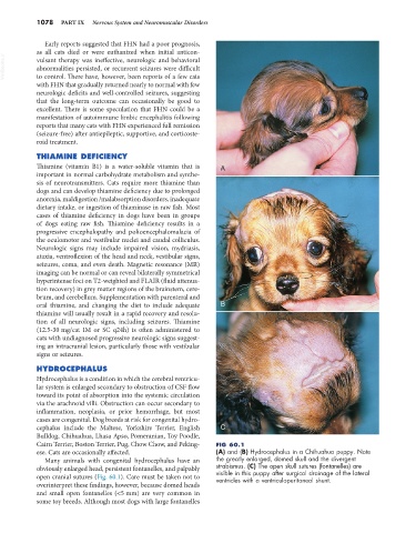
Cairn Terrier, Boston Terrier, Pug, Chow Chow, and Peking- FIG 60.1
ese. Cats are occasionally affected. (A) and (B) Hydrocephalus in a Chihuahua puppy. Note
Many animals with congenital hydrocephalus have an the greatly enlarged, domed skull and the divergent
obviously enlarged head, persistent fontanelles, and palpably strabismus. (C) The open skull sutures (fontanelles) are
visible in this puppy after surgical drainage of the lateral
open cranial sutures (Fig. 60.1). Care must be taken not to ventricles with a ventriculoperitoneal shunt.
overinterpret these findings, however, because domed heads
and small open fontanelles (<5 mm) are very common in
some toy breeds. Although most dogs with large fontanelles