
Page 1100 - Small Animal Internal Medicine, 6th Edition
P. 1100
1072 PART IX Nervous System and Neuromuscular Disorders
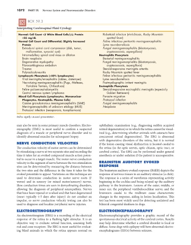
BOX 59.3
VetBooks.ir Interpreting Cerebrospinal Fluid Cytology Rickettsial infection (ehrlichiosis, Rocky Mountain
Normal: Cell Count <5 White Blood Cells/µL; Protein
<25 mg/dL spotted fever)
Normal Cell Count and Differential; Slightly Increased Feline infectious peritonitis meningoencephalitis
Protein Lyme neuroborreliosis
Extradural spinal cord compression (disk, tumor, Fungal meningoencephalitis (blastomycosis,
malformation, synovial cyst) cryptococcosis, aspergillosis)
Intramedullary spinal cord mass or dilation Neutrophilic Pleocytosis
Brain neoplasia Bacterial meningoencephalitis
Degenerative myelopathy Fungal meningoencephalitis (blastomycosis,
Fibrocartilaginous embolism cryptococcosis, aspergillosis)
Trauma Steroid-responsive meningitis arteritis
Polyradiculoneuritis Rocky Mountain spotted fever
Lymphocytic Pleocytosis (>50% lymphocytes) Feline infectious peritonitis meningoencephalitis
Viral meningitis/encephalitis (rabies, distemper) Lyme neuroborreliosis
Necrotizing meningoencephalitis (Pugs, Malteses, Postmyelographic irritant meningitis
Yorkshire Terriers, Chihuahuas) Eosinophilic Pleocytosis
Feline polioencephalomyelitis Steroid-responsive eosinophilic meningitis (especially
Central nervous system lymphoma Golden Retrievers)
Mixed Cell Pleocytosis (Lymphocytes, Mononuclear Parasite migration
Phagocytes, Neutrophils, Plasma Cells) Protozoal infection
Canine granulomatous meningoencephalitis (GME) Fungal meningoencephalitis
Meningoencephalitis of unknown etiology (MUE) Neoplasia
Protozoal infection (neosporosis, toxoplasmosis)
Italics signify unusual presentation.
may also be seen in some primary muscle disorders. Electro- ophthalmic examination (e.g., diagnosing sudden acquired
myography (EMG) is most useful to confirm a suspected retinal degeneration) or in which the retina cannot be visual-
diagnosis of a muscle or peripheral nerve disorder and to ized (e.g., determining whether animals with cataracts have
identify abnormal muscles for subsequent biopsy. concurrent retinal degeneration). The ERG is abnormal
with degenerative disorders of the retina, but it is normal
NERVE CONDUCTION VELOCITIES if the lesion causing visual dysfunction is located caudal to
The conduction velocity of motor nerves can be determined the retina (in the optic nerves, optic chiasm, optic tract, or
by stimulating a nerve at two separate sites and recording the cerebral cortex). The ERG can be performed under general
time it takes for an evoked compound muscle action poten- anesthesia or under sedation if the patient is uncooperative.
tial to occur in a target muscle. The motor nerve conduction
velocity in the segment of nerve between the two stimulation BRAINSTEM AUDITORY EVOKED
sites can be determined by measuring the distance between RESPONSE
the two sites and the difference in the time it takes for the The brainstem auditory evoked response (BAER) depicts the
evoked potentials to appear. Variations on this technique are response of nervous tissues to an auditory stimulus (a click).
used to determine conduction in motor nerve roots (F The response is a series of waveforms representing activity
waves) and in sensory (or mixed motor/sensory) nerves. beginning in the cochlea and being relayed up the auditory
Slow conduction times are seen in demyelinating disorders, pathway in the brainstem. Lesions of the outer, middle, or
allowing the diagnosis of peripheral neuropathies. Nerves inner ear; the peripheral vestibulocochlear nerve; and the
that have been injured or avulsed and that have degenerated brainstem caudal to the midbrain cause characteristic
(onset typically 4-5 days after injury) do not conduct an changes in the response, aiding in lesion localization. This
impulse, so nerve conduction velocity testing can also be test has been most widely used for detecting unilateral and
used to diagnose and localize peripheral nerve injuries. bilateral congenital deafness in dogs.
ELECTRORETINOGRAPHY ELECTROENCEPHALOGRAPHY
An electroretinogram (ERG) is a recording of the electrical Electroencephalography provides a graphic record of the
response of the retina to a flashing light stimulus. It is an spontaneous electrical activity of the cerebral cortex. Results
objective way to evaluate retinal function, assessing both may help determine whether a cerebral disorder is focal or
rod and cone receptors. The ERG is most useful for evaluat- diffuse. Some dogs with epilepsy will have abnormal electro-
ing blind animals in which the retina appears normal on encephalograms (EEGs) between seizures.