
Page 1098 - Small Animal Internal Medicine, 6th Edition
P. 1098
1070 PART IX Nervous System and Neuromuscular Disorders
draw an imaginary line at their most cranial aspect. If that containing ethylenediaminetetraacetic acid (EDTA) to
imaginary line is not perpendicular to the table, then padding prevent clotting but samples for bacterial culture should be
VetBooks.ir should be placed under the animal’s shoulder to bring the free of anticoagulant. The amount of CSF collected ranges
from 0.5 to 3 mL depending on the size of the animal (no
wings into alignment.
The examiner can then use the left index finger to palpate
vein compression may hasten flow but will transiently
the external occipital protuberance and draw a second imag- more than 1 mL/5 kg body weight). Simultaneous jugular
inary line caudally from that site along the dorsal midline. increase intracranial pressure. Blood in the CSF may be the
The needle should be inserted where the two imaginary lines result of the disease or the tap. If it is caused by the proce-
intersect (Fig. 59.7) (Video 59.1). dure, the amount of blood should decrease as the CSF drips
A 1½ or 3-inch-long (3.75-7.5 cm) styletted spinal needle from the needle. If this occurs, some of the less contaminated
is then directed straight in through the skin (perpendicular fluid should be collected in a second tube for cytologic evalu-
to the spine) and into the underlying tissues, with the bevel ation. Mild CSF contamination with hemorrhage (<500
directed cranially. The needle is advanced 1 to 2 mm at a RBCs/µL) does not alter the CSF protein and leukocyte
time, and the stylette is removed so that the clinician can determinations.
look for CSF. While the right hand is used to remove the
stylette, the thumb and first finger of the left hand, which is Lumbar Puncture
rested against the spine for support, should grasp and stabi- The animal is placed in lateral recumbency with its trunk
lize the hub of the needle. A “pop” may be felt as the dorsal flexed. Foam cushions are placed between its limbs and
AO membrane and the dura mater and arachnoid mater are beneath the lumbar region to achieve true lateral position-
penetrated simultaneously (Fig. 59.8). This is not a reliable ing. The spines of the lumbar vertebrae are palpated; the
sign, however, and the level at which the subarachnoid space first dorsal spinous process that can be palpated cranial to
is reached varies greatly with the breed and individual the ilial crests is L6. Lumbar puncture should be performed
animal. It is often very close to the skin surface in toy breeds at the L4-L5 or the L5-L6 space (ideal) in dogs and the
and some cats. L6-L7 space in cats. To access the L5-L6 space, a 3½-inch
If the needle strikes bone, it should be withdrawn, the (8.75-cm) spinal needle is inserted on midline at the cranial
patient position and landmarks reassessed, and the proce- edge of the dorsal spinal process of L6 and directed ven-
dure repeated. If whole blood appears in the spinal needle, trally into the ligamentum flavum (Fig. 59.9). The needle
the needle should be withdrawn and the procedure repeated is passed in a smooth motion through or alongside the
with another sterile needle. When CSF is observed, the fluid caudal spinal cord and cauda equina into the ventral sub-
should be allowed to drip directly from the needle into a test arachnoid space. The animal’s tail and pelvic limbs may
tube. The clinician should check with the laboratory to deter- twitch when the cord is penetrated. Because CSF flow is
mine the type of tube preferred for collection of CSF. Plastic slower from this site and more likely to be contaminated by
tubes are preferred because cells may adhere to glass tubes. blood, cerebellomedullary collection is usually preferred for
Grossly hemorrhagic CSF should be collected into a tube diagnostic purposes.
Needle CSF in subarachnoid space
Dura mater
Arachnoid
mater Leptomeninges
Pia mater
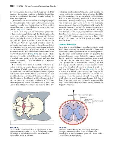
FIG 59.7 FIG 59.8
Landmarks for cerebrospinal fluid (CSF) collection at the Transverse section showing the relationship among the
cerebellomedullary cistern. The site of needle entry is at the meninges, cerebrospinal fluid (CSF), and spinal cord.
intersection of the dorsal midline and the most cranial Needle tip is in the subarachnoid space, as it would be for
aspect of the wings of the atlas. CSF collection or myelography.