
Page 1093 - Small Animal Internal Medicine, 6th Edition
P. 1093
CHAPTER 59 Diagnostic Tests for Nervous System and Neuromuscular Disorders 1065
making diagnostic interpretation of CSF cytology difficult extradural tumors, and dogs with degenerative myelopathy
for at least 1 week after myelography. are most often affected. Fortunately this deterioration is
VetBooks.ir is associated with some risk of complications. The animal is usually transient.
Myelography can be technically difficult to perform and
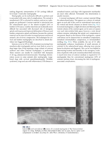
A normal myelogram will show contrast material filling
anesthetized, CSF is collected for analysis, and survey radio-
agent on each side of the cord on ventrodorsal views and in
graphs are performed. Contrast material is introduced into the subarachnoid space. This appears as a column of contrast
the subarachnoid space at the atlanto-occipital (AO) or the ventral and dorsal columns on lateral views (Fig. 59.1).
lumbar (L5/6) space. Lumbar injections are technically more In normal myelograms, a slight elevation and thinning of the
difficult but associated with decreased risk of iatrogenic ventral column of the contrast agent can be seen as it passes
spinal cord trauma and improved delineation of thoracic and over each intervertebral disk space; however, a wide dorsal
lumbar compressive spinal cord lesions, because the contrast column remains, indicating that spinal cord compression is
material can be injected under increased pressure and forced not present. Based on the features of the myelogram, a spinal
around a site of severe compression. The techniques for cer- cord lesion can be characterized as extradural compression,
vical and lumbar myelography are described in detail else- intradural extramedullary compression, or intramedullary
where (see Suggested Readings). swelling (Fig. 59.2). When available, the use of CT after
Seizures occur in 10% to 20% of animals recovering from myelography enhances visualization of small amounts of
anesthesia after myelography and are most likely to occur in contrast in the subarachnoid space, allowing more precise
dogs larger than 29 kg requiring a large volume of contrast lesion localization and diagnosis. This can be very helpful in
medium and when cisternal myelography is performed. determining location and laterality in the emergency evalu-
These seizures can usually be controlled with diazepam ation of patients with acute nonmineralized disk extrusions.
(5-20 mg, administered intravenously). Neurologic deterio- A much lower dose of contrast medium is required for CT
ration occurs in some animals after myelography. Large- myelography than for conventional myelograms (25% of
breed dogs with cervical spondylomyelopathy (Wobbler contrast medium dose), decreasing the risk of myelogram-
syndrome), dogs and cats with inflammatory CNS disease or associated complications.
A B
FIG 59.1
Lateral (A) and ventrodorsal (B) views of a normal myelogram of the thoracolumbar
region in a dog. Multiple calcified intervertebral disks can be seen, but no spinal cord
compression is evident. (Courtesy Dr. John Pharr, University of Saskatchewan.)