
Page 1085 - Small Animal Internal Medicine, 6th Edition
P. 1085
CHAPTER 58 Lesion Localization and the Neurologic Examination 1057
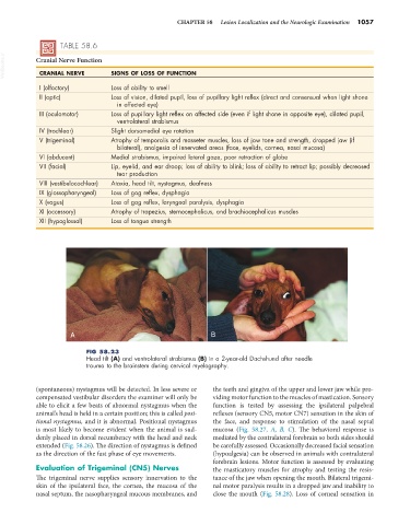
TABLE 58.6
VetBooks.ir Cranial Nerve Function SIGNS OF LOSS OF FUNCTION
CRANIAL NERVE
I (olfactory) Loss of ability to smell
II (optic) Loss of vision, dilated pupil, loss of pupillary light reflex (direct and consensual when light shone
in affected eye)
III (oculomotor) Loss of pupillary light reflex on affected side (even if light shone in opposite eye), dilated pupil,
ventrolateral strabismus
IV (trochlear) Slight dorsomedial eye rotation
V (trigeminal) Atrophy of temporalis and masseter muscles, loss of jaw tone and strength, dropped jaw (if
bilateral), analgesia of innervated areas (face, eyelids, cornea, nasal mucosa)
VI (abducent) Medial strabismus, impaired lateral gaze, poor retraction of globe
VII (facial) Lip, eyelid, and ear droop; loss of ability to blink; loss of ability to retract lip; possibly decreased
tear production
VIII (vestibulocochlear) Ataxia, head tilt, nystagmus, deafness
IX (glossopharyngeal) Loss of gag reflex, dysphagia
X (vagus) Loss of gag reflex, laryngeal paralysis, dysphagia
XI (accessory) Atrophy of trapezius, sternocephalicus, and brachiocephalicus muscles
XII (hypoglossal) Loss of tongue strength
A B
FIG 58.23
Head tilt (A) and ventrolateral strabismus (B) in a 2-year-old Dachshund after needle
trauma to the brainstem during cervical myelography.
(spontaneous) nystagmus will be detected. In less severe or the teeth and gingiva of the upper and lower jaw while pro-
compensated vestibular disorders the examiner will only be viding motor function to the muscles of mastication. Sensory
able to elicit a few beats of abnormal nystagmus when the function is tested by assessing the ipsilateral palpebral
animal’s head is held in a certain position; this is called posi- reflexes (sensory CN5, motor CN7) sensation in the skin of
tional nystagmus, and it is abnormal. Positional nystagmus the face, and response to stimulation of the nasal septal
is most likely to become evident when the animal is sud- mucosa (Fig. 58.27, A, B, C). The behavioral response is
denly placed in dorsal recumbency with the head and neck mediated by the contralateral forebrain so both sides should
extended (Fig. 58.26). The direction of nystagmus is defined be carefully assessed. Occasionally decreased facial sensation
as the direction of the fast phase of eye movements. (hypoalgesia) can be observed in animals with contralateral
forebrain lesions. Motor function is assessed by evaluating
Evaluation of Trigeminal (CN5) Nerves the masticatory muscles for atrophy and testing the resis-
The trigeminal nerve supplies sensory innervation to the tance of the jaw when opening the mouth. Bilateral trigemi-
skin of the ipsilateral face, the cornea, the mucosa of the nal motor paralysis results in a dropped jaw and inability to
nasal septum, the nasopharyngeal mucous membranes, and close the mouth (Fig. 58.28). Loss of corneal sensation in