Page 1083 - Small Animal Internal Medicine, 6th Edition
P. 1083
CHAPTER 58   Lesion Localization and the Neurologic Examination  1055


                                                                 follows the movement. It may be necessary to set up a maze
                   BOX 58.8                                      of objects to assess vision in each eye.
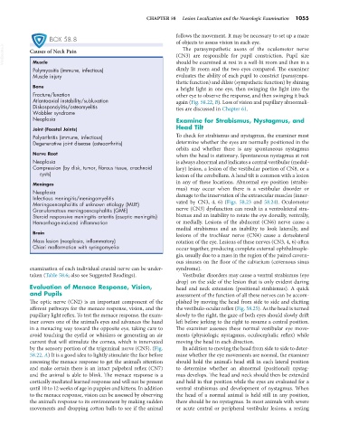
  VetBooks.ir  Causes of Neck Pain                               (CN3) are responsible for pupil constriction. Pupil size
                                                                   The parasympathetic axons of the oculomotor nerve
                                                                 should be examined at rest in a well-lit room and then in a
             Muscle
             Polymyositis (immune, infectious)                   dimly lit room and the two eyes compared. The examiner
             Muscle injury                                       evaluates the ability of each pupil to constrict (parasympa-
                                                                 thetic function) and dilate (sympathetic function) by shining
             Bone                                                a bright light in one eye, then swinging the light into the
             Fracture/luxation                                   other eye to observe the response, and then swinging it back
             Atlantoaxial instability/subluxation                again (Fig. 58.22, B). Loss of vision and pupillary abnormali-
             Diskospondylitis/osteomyelitis                      ties are discussed in Chapter 61.
             Wobbler syndrome
             Neoplasia                                           Examine for Strabismus, Nystagmus, and
             Joint (Facetal Joints)                              Head Tilt
             Polyarthritis (immune, infectious)                  To check for strabismus and nystagmus, the examiner must
             Degenerative joint disease (osteoarthritis)         determine whether the eyes are normally positioned in the
                                                                 orbits and whether there is any spontaneous nystagmus
             Nerve Root                                          when the head is stationary. Spontaneous nystagmus at rest
             Neoplasia                                           is always abnormal and indicates a central vestibular (medul-
             Compression (by disk, tumor, fibrous tissue, arachnoid   lary) lesion, a lesion of the vestibular portion of CN8, or a
               cysts)                                            lesion of the cerebellum. A head tilt is common with a lesion

             Meninges                                            in any of these locations. Abnormal eye position (strabis-
             Neoplasia                                           mus) may occur when there is a vestibular disorder or
             Infectious meningitis/meningomyelitis               damage to the innervation of the extraocular muscles (inner-
             Meningoencephalitis of unknown etiology (MUE)       vated  by  CN3,  4,  6)  (Figs.  58.23  and  58.24).  Oculomotor
             Granulomatous meningoencephalitis (GME)             nerve (CN3) dysfunction can result in a ventrolateral stra-
             Steroid responsive meningitis arteritis (aseptic meningitis)  bismus and an inability to rotate the eye dorsally, ventrally,
             Hemorrhage-induced inflammation                     or medially. Lesions of the abducent (CN6) nerve cause a
                                                                 medial strabismus and an inability to look laterally, and
             Brain                                               lesions of the trochlear nerve (CN4) cause a dorsolateral
             Mass lesion (neoplasia, inflammatory)               rotation of the eye. Lesions of these nerves (CN3, 4, 6) often
             Chiari malformation with syringomyelia              occur together, producing complete external ophthalmople-
                                                                 gia, usually due to a mass in the region of the paired cavern-
                                                                 ous sinuses on the floor of the calvarium (cavernous sinus
            examination of each individual cranial nerve can be under-  syndrome).
            taken (Table 58.6; also see Suggested Readings).       Vestibular disorders may cause a ventral strabismus (eye
                                                                 drop) on the side of the lesion that is only evident during
            Evaluation of Menace Response, Vision,               head and neck extension (positional strabismus). A quick
            and Pupils                                           assessment of the function of all these nerves can be accom-
            The optic nerve (CN2) is an important component of the   plished by moving the head from side to side and eliciting
            afferent pathways for the menace response, vision, and the   the vestibulo-ocular reflex (Fig. 58.25). As the head is turned
            pupillary light reflex. To test the menace response, the exam-  slowly to the right, the gaze of both eyes should slowly drift
            iner covers one of the animal’s eyes and advances the hand   left before jerking to the right to resume a central position.
            in a menacing way toward the opposite eye, taking care to   The examiner assesses these normal vestibular eye move-
            avoid touching the eyelid or whiskers or generating an air   ments (physiologic nystagmus, oculocephalic reflex) while
            current that will stimulate the cornea, which is innervated   moving the head in each direction.
            by the sensory portion of the trigeminal nerve (CN5). (Fig.   In addition to moving the head from side to side to deter-
            58.22, A) It is a good idea to lightly stimulate the face before   mine whether the eye movements are normal, the examiner
            assessing the menace response to get the animal’s attention   should hold the animal’s head still in each lateral position
            and make certain there is an intact palpebral reflex (CN7)   to determine whether an abnormal (positional) nystag-
            and the animal is able to blink. The menace response is a   mus develops. The head and neck should then be extended
            cortically mediated learned response and will not be present   and held in that position while the eyes are evaluated for a
            until 10 to 12 weeks of age in puppies and kittens. In addition   ventral strabismus and development of nystagmus. When
            to the menace response, vision can be assessed by observing   the head of a normal animal is held still in any position,
            the animal’s response to its environment by making sudden   there should be no nystagmus. In most animals with severe
            movements and dropping cotton balls to see if the animal   or acute central or peripheral vestibular lesions, a resting

