
Page 32 - Small Animal Internal Medicine, 6th Edition
P. 32
4 PART I Cardiovascular System Disorders
congenital and acquired abnormalities are more prevalent in It is important to note whether the respiratory difficulty
certain breeds or life stages, or because specific findings are is more intense during a particular phase of respiration. Pro-
VetBooks.ir common in individuals of a given breed (such as a soft left longed, labored inspiration usually is associated with upper
airway obstructive disorders, whereas prolonged expiration
basilar ejection murmur in normal Greyhounds and other
sighthounds).
Physical evaluation of the patient with suspected heart occurs with lower airway obstruction as well as pulmonary
infiltrative disease (including edema). Animals with severely
disease includes observation (for example, of attitude, compromised ventilation may refuse to lie down; rather,
posture, body condition, level of anxiety, respiratory pattern) they stand or sit with elbows abducted to allow maximal
and a general physical examination. The CV examination rib expansion, and they resist being positioned in lateral
itself consists of evaluating the peripheral circulation or dorsal recumbency (orthopnea). Cats with dyspnea
(mucous membranes), systemic veins (especially the jugular often crouch in a sternal position with elbows abducted.
veins), systemic arterial pulses (usually the femoral arteries), Open-mouth breathing usually is a sign of severe respira-
and the precordium (left and right chest wall over the heart), tory distress in cats (Fig. 1.3). The increased respiratory rate
as well as auscultation of the heart and lungs, and palpating associated with excitement, fever, fear, or pain generally can
or percussing for abnormal fluid accumulation (e.g., ascites, be differentiated from dyspnea by careful observation and
subcutaneous edema, pleural effusion). Proficiency in all physical examination.
aspects of the CV examination requires practice but is
important for accurate patient assessment and monitoring. MUCOUS MEMBRANES
Mucous membrane color and capillary refill time (CRT) are
RESPIRATORY PATTERNS used to evaluate peripheral perfusion. The oral mucosa
Respiratory difficulty (dyspnea) usually causes the animal to usually is assessed, although caudal mucous membranes
appear anxious. Increased respiratory effort, flared nostrils, (prepuce or vagina) also can be evaluated. The CRT is deter-
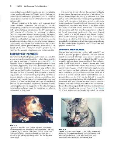
and often a rapid rate of breathing are evident (Fig. 1.2). mined by applying digital pressure to blanch the membrane;
Increased depth of respiration (hyperpnea) can result from color should return within 2 seconds. Slower refill times
hypoxemia, hypercarbia, or acidosis. Pulmonary edema (or occur as a result of dehydration and other causes of decreased
other pulmonary infiltrates) increases lung stiffness; the cardiac output because of high peripheral sympathetic tone
rapid and shallow breathing (tachypnea) that results helps and vasoconstriction. Pale mucous membranes occur with
minimize the work of breathing. In the absence of primary either anemia or peripheral vasoconstriction. The CRT is
lung disease, an increase in resting respiratory rate often is normal in anemic animals unless hypoperfusion also is
an early indicator of pulmonary edema. Lung stiffness also present. However, the CRT can be difficult to assess in
increases with pleural fluid or air accumulation and can severely anemic animals because of the lack of color contrast.
produce tachypnea, too. However, with large-volume pleural In animals with polycythemia (erythrocytosis) or exercise-
effusion or pneumothorax, respiratory motions become induced rear limb weakness, the color of the caudal mem-
increasingly labored and exaggerated as the animal struggles branes should be compared with that of the oral membranes
to expand the collapsed lungs; the respiratory rate often is for evidence of differential cyanosis (see p. 115 in Chapter
not elevated in these cases. 5). If oral membranes are heavily pigmented, the ocular
FIG 1.2
Dyspnea in an older male Golden Retriever with dilated
cardiomyopathy and fulminant pulmonary edema. The dog FIG 1.3
appeared highly anxious, with rapid labored respirations Severe dyspnea is manifested in this cat by open-mouth
and hypersalivation. Respiratory arrest occurred within breathing, infrequent swallowing (drooling saliva), and
minutes of this photograph being taken, but the dog was reluctance to lie down. Note also the dilated pupils
resuscitated. associated with heightened sympathetic tone.