
Page 36 - Small Animal Internal Medicine, 6th Edition
P. 36
8 PART I Cardiovascular System Disorders
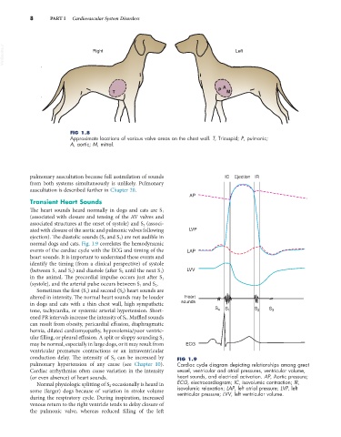
VetBooks.ir Right Left
T P A M
FIG 1.8
Approximate locations of various valve areas on the chest wall. T, Tricuspid; P, pulmonic;
A, aortic; M, mitral.
pulmonary auscultation because full assimilation of sounds IC Ejection IR
from both systems simultaneously is unlikely. Pulmonary
auscultation is described further in Chapter 20.
AP
Transient Heart Sounds
The heart sounds heard normally in dogs and cats are S 1
(associated with closure and tensing of the AV valves and
associated structures at the onset of systole) and S 2 (associ-
ated with closure of the aortic and pulmonic valves following LVP
ejection). The diastolic sounds (S 3 and S 4 ) are not audible in
normal dogs and cats. Fig. 1.9 correlates the hemodynamic
events of the cardiac cycle with the ECG and timing of the LAP
heart sounds. It is important to understand these events and
identify the timing (from a clinical perspective) of systole
(between S 1 and S 2 ) and diastole (after S 2 until the next S 1 ) LVV
in the animal. The precordial impulse occurs just after S 1
(systole), and the arterial pulse occurs between S 1 and S 2 .
Sometimes the first (S 1 ) and second (S 2 ) heart sounds are
altered in intensity. The normal heart sounds may be louder Heart
in dogs and cats with a thin chest wall, high sympathetic sounds
tone, tachycardia, or systemic arterial hypertension. Short- S 4 S 1 S 2 S 3
ened PR intervals increase the intensity of S 1 . Muffled sounds
can result from obesity, pericardial effusion, diaphragmatic
hernia, dilated cardiomyopathy, hypovolemia/poor ventric-
ular filling, or pleural effusion. A split or sloppy-sounding S 1
may be normal, especially in large dogs, or it may result from ECG
ventricular premature contractions or an intraventricular
conduction delay. The intensity of S 2 can be increased by FIG 1.9
pulmonary hypertension of any cause (see Chapter 10). Cardiac cycle diagram depicting relationships among great
Cardiac arrhythmias often cause variation in the intensity vessel, ventricular and atrial pressures, ventricular volume,
(or even absence) of heart sounds. heart sounds, and electrical activation. AP, Aortic pressure;
Normal physiologic splitting of S 2 occasionally is heard in ECG, electrocardiogram; IC, isovolumic contraction; IR,
some (larger) dogs because of variation in stroke volume isovolumic relaxation; LAP, left atrial pressure; LVP, left
ventricular pressure; LVV, left ventricular volume.
during the respiratory cycle. During inspiration, increased
venous return to the right ventricle tends to delay closure of
the pulmonic valve, whereas reduced filling of the left