
Page 35 - Small Animal Internal Medicine, 6th Edition
P. 35
CHAPTER 1 Clinical Manifestations of Cardiac Disease 7
VetBooks.ir
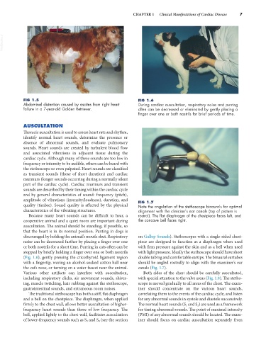
FIG 1.5 FIG 1.6
Abdominal distention caused by ascites from right heart During cardiac auscultation, respiratory noise and purring
failure in a 7-year-old Golden Retriever. often can be decreased or eliminated by gently placing a
finger over one or both nostrils for brief periods of time.
AUSCULTATION
Thoracic auscultation is used to assess heart rate and rhythm,
identify normal heart sounds, determine the presence or
absence of abnormal sounds, and evaluate pulmonary
sounds. Heart sounds are created by turbulent blood flow
and associated vibrations in adjacent tissue during the
cardiac cycle. Although many of these sounds are too low in
frequency or intensity to be audible, others can be heard with
the stethoscope or even palpated. Heart sounds are classified
as transient sounds (those of short duration) and cardiac
murmurs (longer sounds occurring during a normally silent
part of the cardiac cycle). Cardiac murmurs and transient
sounds are described by their timing within the cardiac cycle
and by general characteristics of sound: frequency (pitch),
amplitude of vibrations (intensity/loudness), duration, and FIG 1.7
quality (timbre). Sound quality is affected by the physical Note the angulation of the stethoscope binaurals for optimal
characteristics of the vibrating structures. alignment with the clinician’s ear canals (top of picture is
Because many heart sounds can be difficult to hear, a rostral). The flat diaphragm of the chestpiece faces left, and
cooperative animal and a quiet room are important during the concave bell faces right.
auscultation. The animal should be standing, if possible, so
that the heart is in its normal position. Panting in dogs is
discouraged by holding the animal’s mouth shut. Respiratory on Gallop Sounds). Stethoscopes with a single-sided chest-
noise can be decreased further by placing a finger over one piece are designed to function as a diaphragm when used
or both nostrils for a short time. Purring in cats often can be with firm pressure against the skin and as a bell when used
stopped by briefly holding a finger over one or both nostrils with light pressure. Ideally the stethoscope should have short
(Fig. 1.6), gently pressing the cricothyroid ligament region double tubing and comfortable eartips. The binaural eartubes
with a fingertip, waving an alcohol-soaked cotton ball near should be angled rostrally to align with the examiner’s ear
the cat’s nose, or turning on a water faucet near the animal. canals (Fig. 1.7).
Various other artifacts can interfere with auscultation, Both sides of the chest should be carefully auscultated,
including respiratory clicks, air movement sounds, shiver- with special attention to the valve areas (Fig. 1.8). The stetho-
ing, muscle twitching, hair rubbing against the stethoscope, scope is moved gradually to all areas of the chest. The exam-
gastrointestinal sounds, and extraneous room noises. iner should concentrate on the various heart sounds,
The traditional stethoscope has both a stiff, flat diaphragm correlating them to the events of the cardiac cycle, and listen
and a bell on the chestpiece. The diaphragm, when applied for any abnormal sounds in systole and diastole successively.
firmly to the chest wall, allows better auscultation of higher- The normal heart sounds (S 1 and S 2 ) are used as a framework
frequency heart sounds than those of low frequency. The for timing abnormal sounds. The point of maximal intensity
bell, applied lightly to the chest wall, facilitates auscultation (PMI) of any abnormal sounds should be located. The exam-
of lower-frequency sounds such as S 3 and S 4 (see the section iner should focus on cardiac auscultation separately from