
Page 34 - Small Animal Internal Medicine, 6th Edition
P. 34
6 PART I Cardiovascular System Disorders
BOX 1.5 the area of the femoral triangle, where the femoral artery
enters the leg.
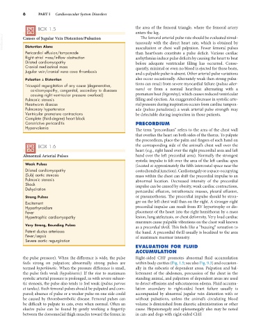
VetBooks.ir Causes of Jugular Vein Distention/Pulsation taneously with the direct heart rate, which is obtained by
The femoral arterial pulse rate should be evaluated simul-
Distention Alone
Pericardial effusion/tamponade auscultation or chest wall palpation. Fewer femoral pulses
than heartbeats constitute a pulse deficit. Various cardiac
Right atrial mass/inflow obstruction arrhythmias induce pulse deficits by causing the heart to beat
Dilated cardiomyopathy before adequate ventricular filling has occurred. Conse-
Cranial mediastinal mass quently, minimal or even no blood is ejected for those beats,
Jugular vein/cranial vena cava thrombosis and a palpable pulse is absent. Other arterial pulse variations
Pulsation ± Distention also occur occasionally. Alternately weak then strong pulsa-
Tricuspid regurgitation of any cause (degenerative, tions can result from severe myocardial failure (pulsus alter-
cardiomyopathy, congenital, secondary to diseases nans) or from a normal heartbeat alternating with a
causing right ventricular pressure overload) premature beat (bigeminy), which causes reduced ventricular
Pulmonic stenosis filling and ejection. An exaggerated decrease in systolic arte-
Heartworm disease rial pressure during inspiration occurs from cardiac tampon-
Pulmonary hypertension ade (pulsus paradoxus); a weak arterial pulse strength may
Ventricular premature contractions be detectable during inspiration in those patients.
Complete (third-degree) heart block
Constrictive pericarditis PRECORDIUM
Hypervolemia The term “precordium” refers to the area of the chest wall
that overlies the heart on both sides of the thorax. To palpate
the precordium, place the palm and fingers of each hand on
BOX 1.6 the corresponding side of the animal’s chest wall over the
heart (e.g., right hand over the right precordial area and left
Abnormal Arterial Pulses hand over the left precordial area). Normally the strongest
systolic impulse is felt over the area of the left cardiac apex
Weak Pulses (located at approximately the fifth intercostal space near the
Dilated cardiomyopathy costochondral junction). Cardiomegaly or a space-occupying
(Sub) aortic stenosis mass within the chest can shift the precordial impulse to an
Pulmonic stenosis abnormal location. Decreased intensity of the precordial
Shock impulse can be caused by obesity, weak cardiac contractions,
Dehydration
pericardial effusion, intrathoracic masses, pleural effusion,
Strong Pulses or pneumothorax. The precordial impulse should be stron-
Excitement ger on the left chest wall than on the right. A stronger right
Hyperthyroidism precordial impulse can result from RV hypertrophy or dis-
Fever placement of the heart into the right hemithorax by a mass
Hypertrophic cardiomyopathy lesion, lung atelectasis, or chest deformity. Very loud cardiac
murmurs cause palpable vibrations on the chest wall known
Very Strong, Bounding Pulses as a precordial thrill. This feels like a “buzzing” sensation to
Patent ductus arteriosus the hand. A precordial thrill usually is localized to the area
Fever/sepsis of maximum murmur intensity.
Severe aortic regurgitation
EVALUATION FOR FLUID
ACCUMULATION
the pulse pressure). When the difference is wide, the pulse Right-sided CHF promotes abnormal fluid accumulation
feels strong on palpation; abnormally strong pulses are within body cavities (Fig. 1.5; see also Fig. 9.3) and occasion-
termed hyperkinetic. When the pressure difference is small, ally in the subcutis of dependent areas. Palpation and bal-
the pulse feels weak (hypokinetic). If the rise to maximum lottement of the abdomen, percussion of the chest in the
systolic arterial pressure is prolonged, as with severe subaor- standing animal, and palpation of dependent areas are used
tic stenosis, the pulse also tends to feel weak (pulsus parvus to detect effusions and subcutaneous edema. Fluid accumu-
et tardus). Both femoral pulses should be palpated and com- lation secondary to right-sided heart failure usually is
pared; absence of pulse or a weaker pulse on one side could accompanied by abnormal jugular vein distention with or
be caused by thromboembolic disease. Femoral pulses can without pulsations, unless the animal’s circulating blood
be difficult to palpate in cats, even when normal. Often an volume is diminished from diuretic administration or other
elusive pulse can be found by gently working a fingertip cause. Hepatomegaly and splenomegaly also may be noted
between the dorsomedial thigh muscles toward the femur, in in cats and dogs with right-sided CHF.