
Page 43 - Small Animal Internal Medicine, 6th Edition
P. 43
CHAPTER 2 Diagnostic Tests for the Cardiovascular System 15
vertebra. The maximum perpendicular short axis is mea-
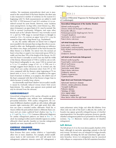
sured in the central third of the heart shadow; the short axis BOX 2.1
VetBooks.ir is also measured in number of vertebrae (to the nearest 0.1) Common Differential Diagnoses for Radiographic Signs
beginning with T4. Both measurements are added to yield
of Cardiomegaly
the VHS. A VHS between 8.5 and 10.5 vertebrae (v) is con-
sidered normal for most breeds. However, some variation Generalized Enlargement of the Cardiac Shadow
exists among breeds. In dogs with a short thorax (e.g., Min- Dilated cardiomyopathy
iature Schnauzer), an upper limit of 11 v may be normal. The Chronic mitral and tricuspid insufficiency
VHS in normal Greyhounds, Whippets, and some other Pericardial effusion
breeds such as the Labrador Retriever may normally exceed Peritoneopericardial diaphragmatic hernia
11 v, and the VHS range in normal Boxers is thought to Tricuspid dysplasia
extend to 12.6 v. In contrast, an upper limit of 9.5 v may be Ventricular or atrial septal defect
normal in dogs with a long thorax (e.g., Dachshund). Patent ductus arteriosus
In cats, the cardiac silhouette on lateral view is aligned Left Atrial Enlargement Alone
more parallel to the sternum than in dogs; this often is accen-
tuated in older cats. Radiographic positioning can influence Early mitral insufficiency
Hypertrophic cardiomyopathy
the relative size, shape, and position of the heart because the Early dilated cardiomyopathy (especially in Doberman
feline thorax is so flexible. On lateral view, the normal cat Pinschers)
heart is less than or equal to two intercostal spaces (ICSs) in (Sub)aortic stenosis
width and less than 70% of the height of the thorax. On DV
view the heart is normally no more than one half the width Left Atrial and Ventricular Enlargement
of the thorax. Measurement of VHS is useful in cats as well. Dilated cardiomyopathy
From lateral radiographs in cats, mean VHS in normal cats Hypertrophic cardiomyopathy
is 7.3 to 7.5 vertebrae (range 6.7-8.1 v). A VHS over 9 v Mitral insufficiency
strongly suggests heart disease in cats. In normal cats, the Aortic insufficiency
mean short-axis cardiac dimension taken from DV or VD Ventricular septal defect
view, compared with the thoracic spine beginning at T4 on Patent ductus arteriosus
(Sub)aortic stenosis
lateral view, is 3.4 to 3.5 v, with 4 v identified as the upper Systemic hypertension
limit of normal. In kittens, as in puppies, the relative size of Hyperthyroidism
the heart compared with that of the thorax is larger than in
adults because of smaller lung volume. Right Atrial and Ventricular Enlargement
An abnormally small heart shadow (microcardia) usually Advanced heartworm disease
is caused by markedly reduced venous return from severe Chronic, severe pulmonary disease
hypovolemia. The cardiac apex appears more pointed and Tricuspid insufficiency
may be elevated from the sternum. Pulmonic stenosis
Tetralogy of Fallot
CARDIOMEGALY Atrial septal defect
Generalized enlargement of the cardiac silhouette on plain Pulmonary hypertension
Mass lesion within the right heart
thoracic radiographs may indicate true cardiomegaly or
pericardial distention. With cardiac enlargement, the con-
tours of different chambers usually are still evident, although
massive right ventricular (RV) and right atrial (RA) dila-
tion can cause a rounded cardiac silhouette. Fluid, fat, or main pulmonary artery bulge, and often RA dilation. Even
viscera within the pericardium tends to obliterate these con- when only one side of the heart is affected, the cardiac sil-
tours and can create a globoid heart shadow (see Fig. 9.1, houette may appear generally enlarged because of chamber
p. 175 and Fig. 9.4, p. 179). Common differential diagnoses superimposition. For descriptive purposes, however, specific
for cardiac enlargement patterns are listed in Box 2.1. A chamber enlargement patterns are presented in the following
clock-face analogy is often used to identify regions on cardiac sections. Fig. 2.2 illustrates various patterns of chamber
silhouette where specific chamber or vascular enlargement enlargement.
typically are seen, especially on DV/VD view.
Left Atrium
CARDIAC CHAMBER The left atrium (LA) is the most dorsocaudal chamber of the
ENLARGEMENT PATTERNS heart, although its auricular appendage extends to the left
Most diseases that cause cardiac dilation or hypertrophy and craniad. On lateral view, an enlarged LA bulges dorsally
affect two or more chambers. For example, mitral insuffi- and caudally, elevating the left and sometimes right main-
ciency leads to left ventricular (LV) and left atrial (LA) stem bronchus. Severe LA enlargement may be associated
enlargement; pulmonic stenosis causes RV enlargement, a with collapse or compression of the left mainstem bronchus.