
Page 44 - Small Animal Internal Medicine, 6th Edition
P. 44
16 PART I Cardiovascular System Disorders
VetBooks.ir
A B
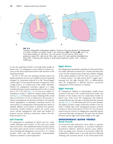
FIG 2.2
Common radiographic enlargement patterns. Diagrams indicating direction of enlargement
of cardiac chambers and great vessels in the dorsoventral (A) and lateral (B) views. Ao,
Aorta (descending); LA, left atrium; LAu, left auricle; LV, left ventricle; MPA, main
pulmonary artery; RA, right atrium; RAu, right auricle; RV, right ventricle. (Modified from
Ware WA: Cardiovascular disease in small animal medicine, London, 2011, Manson
Publishing.)
In cats, the caudal heart border is normally quite straight on Right Atrium
lateral view; LA enlargement causes subtle to marked con- RA enlargement expands the cranial heart border and widens
vexity of the dorsocaudal heart border with elevation of the the cardiac silhouette on lateral view. Tracheal elevation may
mainstem bronchi. occur over the cranial portion of the heart shadow. Bulging
On DV or VD view, the mainstem bronchi tend to be of the cardiac shadow on DV/VD view occurs in the 9- to
displaced laterally and may curve slightly around a markedly 11-o’clock position. The right atrium (RA) is largely super-
enlarged LA (sometimes referred to as the “bowed-legged imposed over the right ventricle (RV), so differentiation
cowboy sign”). Concurrent left auricular enlargement causes from RV enlargement is difficult; however, concurrent
a bulge in the 2- to 3-o’clock area of the cardiac silhouette. enlargement of both chambers is common.
Massive LA enlargement sometimes appears as a large,
rounded soft tissue opacity superimposed over the LV apical Right Ventricle
area on DV (VD) view (Fig. 2.3). In some cats, marked LA RV enlargement (dilation or hypertrophy) usually causes
enlargement creates a “valentine”-shaped cardiac silhouette increased convexity of the cranioventral heart border and
(see Fig. 8.7, p. 169) because it causes widening of the cranial elevation of the trachea over the cranial heart border on
aspect of the heart. LA size is influenced by the pressure or lateral view. With severe RV enlargement and relatively
volume load imposed, as well as by its duration. For example, normal left heart size, the apex is elevated from the sternum
mitral regurgitation of gradually increasing severity can (see Fig. 10.1, p. 191); the carina and CaVC are also elevated.
cause massive LA enlargement without pulmonary edema, if The degree of sternal contact of the heart shadow is not, by
chamber dilation occurs slowly at relatively low pressure. itself, a reliable sign of RV enlargement because of breed
Conversely, chordae tendineae rupture can acutely cause variation in chest conformation. On DV/VD view, the heart
severe valvular regurgitation with rapid and marked LA tends to take on a reverse-D configuration, especially without
pressure increase, leading to pulmonary edema with rela- concurrent left-sided enlargement. The apex may be shifted
tively normal LA size. leftward, and the right heart border bulges to the right.
Left Ventricle INTRATHORACIC BLOOD VESSELS
LV enlargement is manifested on lateral view by a taller Great Vessels
cardiac silhouette with elevation of the carina and caudal The aorta and main pulmonary artery dilate in response
vena cava (CaVC). The caudal heart border becomes convex, to chronic arterial hypertension or increased turbulence
but cardiac apical sternal contact is maintained. On DV/VD (poststenotic dilation). Subaortic stenosis causes dilation
view, rounding and enlargement occur in the 2- to 5-o’clock of the ascending aorta. Because of its location within the
position. Some cats with hypertrophic cardiomyopathy mediastinum, dilation here is not easily detected, although
maintain a pointed LV apical appearance. widening and increased opacity of the dorsocranial heart