
Page 45 - Small Animal Internal Medicine, 6th Edition
P. 45
CHAPTER 2 Diagnostic Tests for the Cardiovascular System 17
VetBooks.ir
A B
FIG 2.3
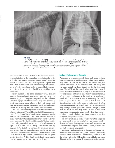
Lateral (A) and dorsoventral (B) views from a dog with chronic mitral regurgitation.
Marked left ventricular and atrial enlargement are evident. Dorsal displacement of the
carina and pulmonary venous distension (arrows) are seen in A; the caudal edge of the
left atrium (arrows), superimposed over the ventricular shadow, and a prominent left
auricular bulge (arrowhead) are seen in B.
shadow may be observed. Patent ductus arteriosus causes a Lobar Pulmonary Vessels
localized dilation in the descending aorta just caudal to the Pulmonary arteries are located dorsal and lateral to their
arch where the ductus exits; this “ductus bump” is seen on accompanying veins and bronchi. In other words, pulmo-
DV or VD view in the 2- to 3-o’clock position. A prominent nary veins are “ventral and central.” On lateral view, the
aortic arch is more common in cats than dogs. The thoracic cranial lobar vessels in the nondependent (“up-side”) lung
aorta of older cats also may have an undulating appear- are more ventral and larger than those in the dependent
ance. Systemic hypertension should be a consideration in lung. The width of the cranial lobar vessels is measured
these cases. where they cross the fourth rib in dogs or at the cranial heart
Severe dilation of the main pulmonary trunk (usually border (fourth to fifth rib) in cats. These vessels are normally
associated with pulmonic stenosis or pulmonary hyperten- 0.5 to 1 times the diameter of the proximal one third of the
sion) can appear as a bulge superimposed over the trachea on fourth rib. The DV view is best for evaluating the caudal
lateral radiograph. On DV view in the dog, main pulmonary pulmonary vessels. The caudal lobar vessels that are 0.5 to 1
trunk enlargement causes a bulge in the 1- to 2-o’clock posi- times the width of the ninth (dogs) or tenth (cats) rib at the
tion. In the cat, the main pulmonary trunk is slightly more point of intersection are normal. However, in many normal
medial and is usually obscured within the mediastinum. dogs, the right caudal pulmonary vessels are slightly wider
The CaVC normally angles cranioventrally from the dia- than the ninth rib. This may be true in cats as well. Four
phragm to the heart. The width of the CaVC is approximately pulmonary vascular patterns are usually described: overcir-
that of the descending thoracic aorta, although its size culation, undercirculation, prominent pulmonary arteries,
changes with respiration. The CaVC-cardiac junction is and prominent pulmonary veins.
pushed dorsally with enlargement of either ventricle. Persis- An overcirculation pattern occurs when the lungs are
tent widening of the CaVC could indicate RV failure, cardiac hyperperfused, as occurs with left-to-right shunts, overhy-
tamponade, pericardial constriction, or other obstruction to dration, and other hyperdynamic states. Pulmonary arteries
right heart inflow. The following comparative findings and veins are both prominent. The increased perfusion also
suggest CaVC distention: CaVC/aortic diameter (at same generally increases lung opacity.
ICS) greater than 1.5; CaVC/length of the thoracic vertebra Pulmonary undercirculation is characterized by thin pul-
directly above the tracheal bifurcation greater than 1.3; and monary arteries and veins, along with increased pulmonary
CaVC/width of right fourth rib (just ventral to the spine) lucency. Severe dehydration, hypovolemia, obstruction to
greater than 3.5. A thin CaVC can indicate hypovolemia, RV inflow, right-sided CHF, and tetralogy of Fallot can cause
poor venous return, or pulmonary overinflation. this pattern. Some animals with pulmonic stenosis appear to