
Page 49 - Small Animal Internal Medicine, 6th Edition
P. 49
CHAPTER 2 Diagnostic Tests for the Cardiovascular System 21
Long-axis 4-chamber view 4-chamber (inflow) view
VetBooks.ir
RV
VS TV RA
LV RV LV
CH
PM MV LA
LVW RA
LA
AS
Long-axis LV outflow view
5-chamber (LV outflow) view
RV
RA
AO
LV
LC
LA
RPA RV LV
RA
AO
LA
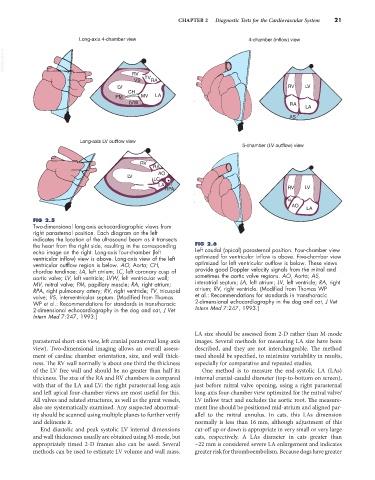
FIG 2.5
Two-dimensional long-axis echocardiographic views from
right parasternal position. Each diagram on the left
indicates the location of the ultrasound beam as it transects
the heart from the right side, resulting in the corresponding FIG 2.6
echo image on the right. Long-axis four-chamber (left Left caudal (apical) parasternal position. Four-chamber view
ventricular inflow) view is above. Long-axis view of the left optimized for ventricular inflow is above. Five-chamber view
ventricular outflow region is below. AO, Aorta; CH, optimized for left ventricular outflow is below. These views
chordae tendinae; LA, left atrium; LC, left coronary cusp of provide good Doppler velocity signals from the mitral and
aortic valve; LV, left ventricle; LVW, left ventricular wall; sometimes the aortic valve regions. AO, Aorta; AS,
MV, mitral valve; PM, papillary muscle; RA, right atrium; interatrial septum; LA, left atrium; LV, left ventricle; RA, right
RPA, right pulmonary artery; RV, right ventricle; TV, tricuspid atrium; RV, right ventricle. (Modified from Thomas WP
valve; VS, interventricular septum. (Modified from Thomas et al.: Recommendations for standards in transthoracic
WP et al.: Recommendations for standards in transthoracic 2-dimensional echocardiography in the dog and cat, J Vet
2-dimensional echocardiography in the dog and cat, J Vet Intern Med 7:247, 1993.)
Intern Med 7:247, 1993.)
LA size should be assessed from 2-D rather than M-mode
parasternal short-axis view, left cranial parasternal long-axis images. Several methods for measuring LA size have been
view). Two-dimensional imaging allows an overall assess- described, and they are not interchangeable. The method
ment of cardiac chamber orientation, size, and wall thick- used should be specified, to minimize variability in results,
ness. The RV wall normally is about one third the thickness especially for comparative and repeated studies.
of the LV free wall and should be no greater than half its One method is to measure the end-systolic LA (LAs)
thickness. The size of the RA and RV chambers is compared internal cranial-caudal diameter (top-to-bottom on screen),
with that of the LA and LV; the right parasternal long axis just before mitral valve opening, using a right parasternal
and left apical four-chamber views are most useful for this. long-axis four-chamber view optimized for the mitral valve/
All valves and related structures, as well as the great vessels, LV inflow tract and excludes the aortic root. The measure-
also are systematically examined. Any suspected abnormal- ment line should be positioned mid-atrium and aligned par-
ity should be scanned using multiple planes to further verify allel to the mitral annulus. In cats, this LAs dimension
and delineate it. normally is less than 16 mm, although adjustment of this
End diastolic and peak systolic LV internal dimensions cut-off up or down is appropriate in very small or very large
and wall thicknesses usually are obtained using M-mode, but cats, respectively. A LAs diameter in cats greater than
appropriately timed 2-D frames also can be used. Several ~22 mm is considered severe LA enlargement and indicates
methods can be used to estimate LV volume and wall mass. greater risk for thromboembolism. Because dogs have greater