
Page 52 - Small Animal Internal Medicine, 6th Edition
P. 52
24 PART I Cardiovascular System Disorders
VetBooks.ir T
TW
RVW
RV
IVS
AV AO
LV
AMV
PMV
LA
LVW
3
1 2
A B
C C D
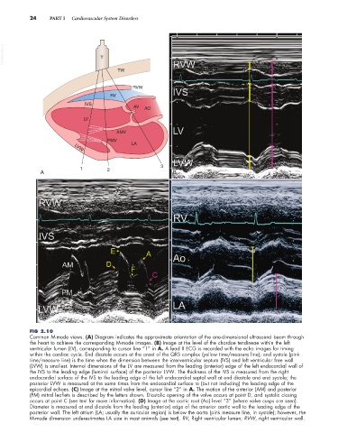
FIG 2.10
Common M-mode views. (A) Diagram indicates the approximate orientation of the one-dimensional ultrasound beam through
the heart to achieve the corresponding M-mode images. (B) Image at the level of the chordae tendineae within the left
ventricular lumen (LV), corresponding to cursor line “1” in A. A lead II ECG is recorded with the echo images for timing
within the cardiac cycle. End diastole occurs at the onset of the QRS complex (yellow time/measure line); end systole (pink
time/measure line) is the time when the dimension between the interventricular septum (IVS) and left ventricular free wall
(LVW) is smallest. Internal dimensions of the LV are measured from the leading (anterior) edge of the left endocardial wall of
the IVS to the leading edge (luminal surface) of the posterior LVW. The thickness of the IVS is measured from the right
endocardial surface of the IVS to the leading edge of the left endocardial septal wall at end diastole and end systole; the
posterior LVW is measured at the same times from the endocardial surface to (but not including) the leading edge of the
epicardial echoes. (C) Image at the mitral valve level, cursor line “2” in A. The motion of the anterior (AM) and posterior
(PM) mitral leaflets is described by the letters shown. Diastolic opening of the valve occurs at point D, and systolic closing
occurs at point C (see text for more information). (D) Image at the aortic root (Ao) level “3” (where valve cusps are seen).
Diameter is measured at end diastole from the leading (anterior) edge of the anterior aortic wall to the leading edge of the
posterior wall. The left atrium (LA; usually the auricular region) is below the aorta (pink measure line, in systole); however, the
M-mode dimension underestimates LA size in most animals (see text). RV, Right ventricular lumen; RVW, right ventricular wall.