
Page 53 - Small Animal Internal Medicine, 6th Edition
P. 53
CHAPTER 2 Diagnostic Tests for the Cardiovascular System 25
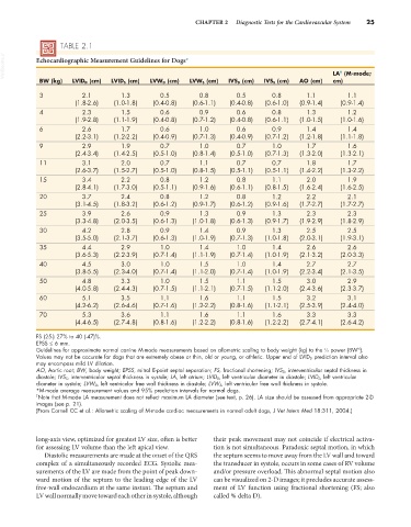
TABLE 2.1
VetBooks.ir Echocardiographic Measurement Guidelines for Dogs* LA (M-mode;
†
BW (kg) LVID D (cm) LVID S (cm) LVW D (cm) LVW S (cm) IVS D (cm) IVS S (cm) AO (cm) cm)
3 2.1 1.3 0.5 0.8 0.5 0.8 1.1 1.1
(1.8-2.6) (1.0-1.8) (0.4-0.8) (0.6-1.1) (0.4-0.8) (0.6-1.0) (0.9-1.4) (0.9-1.4)
4 2.3 1.5 0.6 0.9 0.6 0.8 1.3 1.2
(1.9-2.8) (1.1-1.9) (0.4-0.8) (0.7-1.2) (0.4-0.8) (0.6-1.1) (1.0-1.5) (1.0-1.6)
6 2.6 1.7 0.6 1.0 0.6 0.9 1.4 1.4
(2.2-3.1) (1.2-2.2) (0.4-0.9) (0.7-1.3) (0.4-0.9) (0.7-1.2) (1.2-1.8) (1.1-1.8)
9 2.9 1.9 0.7 1.0 0.7 1.0 1.7 1.6
(2.4-3.4) (1.4-2.5) (0.5-1.0) (0.8-1.4) (0.5-1.0) (0.7-1.3) (1.3-2.0) (1.3-2.1)
11 3.1 2.0 0.7 1.1 0.7 0.7 1.8 1.7
(2.6-3.7) (1.5-2.7) (0.5-1.0) (0.8-1.5) (0.5-1.1) (0.5-1.1) (1.4-2.2) (1.3-2.2)
15 3.4 2.2 0.8 1.2 0.8 1.1 2.0 1.9
(2.8-4.1) (1.7-3.0) (0.5-1.1) (0.9-1.6) (0.6-1.1) (0.8-1.5) (1.6-2.4) (1.6-2.5)
20 3.7 2.4 0.8 1.2 0.8 1.2 2.2 2.1
(3.1-4.5) (1.8-3.2) (0.6-1.2) (0.9-1.7) (0.6-1.2) (0.9-1.6) (1.7-2.7) (1.7-2.7)
25 3.9 2.6 0.9 1.3 0.9 1.3 2.3 2.3
(3.3-4.8) (2.0-3.5) (0.6-1.3) (1.0-1.8) (0.6-1.3) (0.9-1.7) (1.9-2.9) (1.8-2.9)
30 4.2 2.8 0.9 1.4 0.9 1.3 2.5 2.5
(3.5-5.0) (2.1-3.7) (0.6-1.3) (1.0-1.9) (0.7-1.3) (1.0-1.8) (2.0-3.1) (1.9-3.1)
35 4.4 2.9 1.0 1.4 1.0 1.4 2.6 2.6
(3.6-5.3) (2.2-3.9) (0.7-1.4) (1.1-1.9) (0.7-1.4) (1.0-1.9) (2.1-3.2) (2.0-3.3)
40 4.5 3.0 1.0 1.5 1.0 1.4 2.7 2.7
(3.8-5.5) (2.3-4.0) (0.7-1.4) (1.1-2.0) (0.7-1.4) (1.0-1.9) (2.2-3.4) (2.1-3.5)
50 4.8 3.3 1.0 1.5 1.1 1.5 3.0 2.9
(4.0-5.8) (2.4-4.3) (0.7-1.5) (1.1-2.1) (0.7-1.5) (1.1-2.0) (2.4-3.6) (2.3-3.7)
60 5.1 3.5 1.1 1.6 1.1 1.5 3.2 3.1
(4.2-6.2) (2.6-4.6) (0.7-1.6) (1.2-2.2) (0.8-1.6) (1.1-2.1) (2.5-3.9) (2.4-4.0)
70 5.3 3.6 1.1 1.6 1.1 1.6 3.3 3.3
(4.4-6.5) (2.7-4.8) (0.8-1.6) (1.2-2.2) (0.8-1.6) (1.2-2.2) (2.7-4.1) (2.6-4.2)
FS (25-) 27% to 40 (-47)%.
EPSS ≤ 6 mm.
Guidelines for approximate normal canine M-mode measurements based on allometric scaling to body weight (kg) to the ⅓ power (BW ).
⅓
Values may not be accurate for dogs that are extremely obese or thin, old or young, or athletic. Upper end of LVID D prediction interval also
may encompass mild LV dilation.
AO, Aortic root; BW, body weight; EPSS, mitral E-point septal separation; FS, fractional shortening; IVS D, interventricular septal thickness in
diastole; IVS S , interventricular septal thickness in systole; LA, left atrium; LVID D , left ventricular diameter in diastole; LVID S, left ventricular
diameter in systole; LVW D, left ventricular free wall thickness in diastole; LVW S , left ventricular free wall thickness in systole.
*M-mode average measurement values and 95% prediction intervals for normal dogs.
† Note that M-mode LA measurement does not reflect maximum LA diameter (see text, p. 26). LA size should be assessed from appropriate 2-D
images (see p. 21).
(From Cornell CC et al.: Allometric scaling of M-mode cardiac measurements in normal adult dogs, J Vet Intern Med 18:311, 2004.)
long-axis view, optimized for greatest LV size, often is better their peak movement may not coincide if electrical activa-
for assessing LV volume than the left apical view. tion is not simultaneous. Paradoxic septal motion, in which
Diastolic measurements are made at the onset of the QRS the septum seems to move away from the LV wall and toward
complex of a simultaneously recorded ECG. Systolic mea- the transducer in systole, occurs in some cases of RV volume
surements of the LV are made from the point of peak down- and/or pressure overload. This abnormal septal motion also
ward motion of the septum to the leading edge of the LV can be visualized on 2-D images; it precludes accurate assess-
free-wall endocardium at the same instant. The septum and ment of LV function using fractional shortening (FS; also
LV wall normally move toward each other in systole, although called % delta D).