
Page 51 - Small Animal Internal Medicine, 6th Edition
P. 51
CHAPTER 2 Diagnostic Tests for the Cardiovascular System 23
Long-axis view 1 for ventricular measurements; however, it may be more dif-
ficult to verify that the beam is transecting the LV at its
VetBooks.ir RVO PV maximum dimension. Precise positioning of the ultrasound
beam within the heart (perpendicular to the structures to
RC
LV
for accurate M-mode measurements and calculations. For
NC AO be measured) and clear endocardial images are essential
LA example, papillary muscles within the LV must be avoided
when measuring free-wall thickness. Fig. 2.10 illustrates
standard M-mode views. In cases where the M-mode cursor
cannot be optimally aligned (e.g., in animals with focal or
Long-axis view 2 asymmetric hypertrophy), wall thickness measurements
from 2-D images are preferred, although in animals with
a high heart rate, obtaining true end-diastolic frames can
be uncertain.
RV
LV
Common Measurements and
C a RA Normal Values
V C RAu
The standard dimensions measured with M-mode and their
timing also are indicated in Fig. 2.10. The “leading edge”
technique is used when possible (i.e., from the edge closest
to the transducer [leading edge] of one side of the dimension
Long-axis view 3 to the leading edge of the other). In this way, only one endo-
cardial thickness is included in the measurement. LV wall
RVO and interventricular septal thicknesses, as well as LV chamber
PV dimensions, should be determined at the level of the chordae
LV PA tendineae rather than the apex or mitral valve level. Mea-
surements also may be taken from 2-D images if they are of
LA high resolution, and frames from the appropriate times in
the cardiac cycle are used. Body size greatly influences echo-
cardiographic measurements, especially in dogs. However,
the relationship between body weight or surface area and
cardiac dimensions is not linear. Rather, cardiac linear
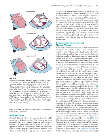
FIG 2.9
Left cranial parasternal long-axis views optimized for aortic dimensions are more closely related to body length, which is
⅓
root (above), right atrium and auricle (middle), and right proportional to body weight to the ⅓ power (BW ). Allome-
ventricular outflow and main pulmonary artery (below). tric scaling has been used to generate guidelines for common
These views are used to evaluate the heart base and can cardiac dimension measurements in normal dogs (see Table
provide good Doppler signals for tricuspid and pulmonary 2.1). Mean values are listed for selected weights along with
flows. AO, Aorta; CaVC, caudal vena cava; LA, left atrium; 95% prediction intervals. However, these prediction inter-
LV, left ventricle; PA, pulmonary artery; PV, pulmonary
valve; RA, right atrium; RAu, right auricle; RC, NC, right vals are quite wide, especially for larger dogs, and may
and noncoronary cusps of aortic valve; RV, right ventricle; encompass some degree of LV enlargement. Somatotype and
RVO, right ventricular outflow tract. (Modified from Thomas breed may have additional influence on normal echo values
WP et al.: Recommendations for standards in transthoracic in some dogs. For example, healthy Boxers can have increased
2-dimensional echocardiography in the dog and cat, J Vet LV wall thickness and smaller aortic dimensions relative to
Intern Med 7:247, 1993.) nonBoxer dogs, although chamber dimensions are compa-
rable. Slightly higher LV wall thickness and chamber dimen-
sions have been observed in Greyhounds compared with
beam placement for standard measurements and calcula- other dogs of comparable weight. Endurance training also
tions can be a limitation. affects measured parameters, reflecting the increased cardiac
mass and volume associated with frequent and sustained
M-Mode Views strenuous exercise. Normal measurements in cats are more
Standard M-mode views are obtained from the right uniform but also are influenced by body size (Table 2.2).
parasternal transducer position. The M-mode cursor is Chamber volume and ejection fraction are better estimated
positioned with 2-D guidance using the right paraster- from optimized 2-D frames using the modified Simpsons’
nal short-axis view; angling the transducer so that the LV method rather than M-mode images because of the greater
appears as round as possible helps ensure the ultrasound potential for inaccurate geometric assumptions from one-
beam is oriented perpendicular to the axis of the LV. Some dimensional measurements (see Suggested Readings for
clinicians prefer using the long-axis view to obtain images further information). The right parasternal four-chamber