
Page 48 - Small Animal Internal Medicine, 6th Edition
P. 48
20 PART I Cardiovascular System Disorders
(for very large cats); have the owner mix the contents of the abnormalities, are evident; actual blood flow is not visualized
appropriately sized capsule with a tiny amount of wet food with 2-D or M-mode imaging alone.
VetBooks.ir and administer on an empty stomach. If additional sedation Common Two-Dimensional
is required to perform the echo, a light dose of butorphanol
Echocardiographic Views
can be effective. Other strategies have included aceproma-
zine (0.1 mg/kg IM) followed in 15 minutes by ketamine A variety of planes can be imaged from several chest wall
(2 mg/kg [or 5-10 mg/cat] IV), although this can undesir- locations. Most standard views are obtained from either the
ably increase heart rate. right or left parasternal positions (directly over the heart and
close to the sternum). Images sometimes are obtained from
TWO-DIMENSIONAL the subxiphoid (subcostal) position. Long-axis views are
ECHOCARDIOGRAPHY obtained with the imaging plane parallel to the long axis of
Two-dimensional echocardiography displays a plane of the heart; short-axis views are perpendicular to this plane
tissue (depth and width). Anatomic structure and motion, (Figs. 2.4 to 2.9). Images are described by the location of the
including changes caused by various acquired or congenital transducer and the imaging plane used (for example, right
RV PM
RVD
LVO
LV
PMV AMV
CH
C F D
E
D
C
RV B RV
A TV
LV NC RC PV
PPM LC
LA
APM
B E
RV RA
RAu
LV
AO
CaVC PA
RPA
A F LPA
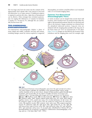
FIG 2.4
Two-dimensional short-axis echocardiographic views from the right parasternal position.
The center diagram indicates the orientation of the ultrasound beam used to image
cardiac structures at the six levels shown. Several of these positions guide M-mode beam
placement, and occasionally can be used for Doppler evaluation of tricuspid and
pulmonary flows. Corresponding echo images are shown clockwise from the bottom.
(A) Apex. (B) Papillary muscle. (C) Chordae tendineae. (D) Mitral valve. (E) Aortic valve.
(F) Pulmonary artery. AMV, Anterior (septal) mitral valve cusp; AO, aorta; APM, anterior
papillary muscle; CaVC, caudal vena cava; CH, chordae tendineae; LA, left atrium; LPA,
left pulmonary artery; LV, left ventricle; LVO, left ventricular outflow tract; PA, pulmonary
artery; PM, papillary muscle; PMV, posterior mitral valve cusp; PPM, posterior papillary
muscle; PV, pulmonary valve; RA, right atrium; RAu, right auricle; RC, LC, NC, right, left,
and noncoronary cusps of aortic valve; RPA, right pulmonary artery; RV, right ventricle;
RVO, right ventricular outflow tract; TV, tricuspid valve. (Modified from Thomas WP et al.:
Recommendations for standards in transthoracic 2-dimensional echocardiography in the
dog and cat, J Vet Intern Med 7:247, 1993.)