
Page 50 - Small Animal Internal Medicine, 6th Edition
P. 50
22 PART I Cardiovascular System Disorders
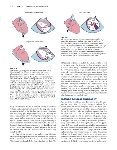
Long-axis 2-chamber view Short-axis view
VetBooks.ir PV
RV
RC LC PA
LV TV NC
AMV
PMV LAu RA
LA
FIG 2.8
Left cranial parasternal short-axis view optimized for right
Long-axis LV outflow view ventricular inflow and outflow. This view is useful for
Doppler interrogation of tricuspid and pulmonary artery
flows. PA, Pulmonary artery; PV, pulmonary valve; RA, right
atrium; RC, LC, NC, right, left, and noncoronary cusps of
aortic valve; RV, right ventricle; TV, tricuspid valve.
R (Modified from Thomas WP et al.: Recommendations for
LV V O standards in transthoracic 2-dimensional echocardiography
RC in the dog and cat, J Vet Intern Med 7:247, 1993.)
NC AO
LA
2-D image is optimized to include the LA and auricle, as well
as the aortic valve. The internal LA dimension is measured
in early diastole, along a line extending from and parallel to
FIG 2.7
Left caudal (apical) parasternal two-dimensional views the commissure formed by the closed left and noncoronary
optimized for left ventricular inflow and left auricle aortic valve cusps. The aortic dimension measurement, from
(2-chamber view; above) and left ventricular outflow the same frozen 2-D frame, also aligns with the same valve
(3-chamber view; below); the outflow view sometimes is commissure and includes only one sinus of Valsalva. The
obtained with the aorta pointing to the lower left of the LAd:aortic root ratio using this short-axis method in normal
image. The 3-chamber view can provide good alignment dogs usually is between 1.3 to 1.4. Ratios over 1.5 to 1.6
with left ventricular outflow velocity (although the subcostal indicate LA enlargement. However, LA size evaluation using
position [not illustrated here] is often better). AMV, Anterior
(septal) mitral valve cusp; AO, aorta; LA, left atrium; LAu, this short-axis method can be confounded by the fact that
left auricle; LV, left ventricle; PMV, posterior mitral valve maximal LA size is not measured, by variability in the
cusp; RC, NC, right and noncoronary cusps of aortic valve; imaging plane used among echocardiographers, and by
RVO, right ventricular outflow tract. (Modified from Thomas inconsistent measurement timing, especially when the aortic
WP et al.: Recommendations for standards in transthoracic valve leaflets are not clearly imaged.
2-dimensional echocardiography in the dog and cat, J Vet
Intern Med 7:247, 1993.) M-MODE ECHOCARDIOGRAPHY
This modality provides a one-dimensional (depth) view
into the heart. M-mode images represent echoes from
body size variation, the LA dimension usually is compared various tissue interfaces along the axis of the beam (dis-
with an aortic measurement, both for the long-axis, systolic played vertically on the screen). These echoes, which move
dimension as well as for the short-axis, diastolic dimension during the cardiac cycle, are displayed against time (on
(see next paragraph). Regarding the long axis, LAs compari- the horizontal axis). Thus the “wavy” lines seen on these
son, some clinicians advocate using the distance between the recordings correspond to the positions of specific struc-
open aortic leaflets (at the valve hinge points) during mid- tures in relation to the transducer, as well as to each other
systole (AoVs), measured from a right long-axis view opti- at any point in time. Accurate M-mode beam placement,
mized for the LV outflow tract and aortic valve. The ratio of using a moveable cursor line superimposed on an appro-
Las:AoVs in normal dogs is thought to be <2.6. However, if priate 2-D (real-time) image, is essential for measurement
the 2-D aortic root diameter is measured across the sinuses purposes. M-mode images usually provide cleaner resolu-
of Valsalva, the ratio of LAs:aortic root in normal dogs tion of cardiac borders than 2-D images because of their
usually is ≤1.9. higher sampling rate. Measurements of cardiac dimensions
Another LA size assessment method, often used in dogs, and motion throughout the cardiac cycle often are more
employs the right parasternal short-axis view and compares accurately obtained from M-mode tracings, especially when
the LA dimension in diastole (LAd) to the aortic diameter. coupled with a simultaneously recorded ECG (or phono-
This sometimes is called the “Scandinavian” method. The cardiogram). Difficulty in achieving consistent and accurate