
Page 548 - Small Animal Internal Medicine, 6th Edition
P. 548
520 PART IV Hepatobiliary and Exocrine Pancreatic Disorders
decreased intravascular oncotic pressure, or altered vascu- rare in cats. It is also recognized in some cases of acute liver
lar permeability and insufficient resorption), singly or in disease. Typically, it results in the triad of ascites, HE, and
VetBooks.ir combination, apply to cats and dogs with hepatobiliary dis- GI congestion, and propensity to ulceration. It is caused by
the increased resistance to blood flow through the sinusoids
eases. In parenchymal liver disease, the commonest cause of
ascites formation is portal hypertension, which is a sustained
the portal vein or caudal vena cava, such as those caused by
increase in pressure in the portal system, with or without of the liver or, less commonly, by more direct obstructions to
a contribution from reduced serum albumin concentration thromboemboli. The presence of a large arteriovenous fistula
(Fig. 33.1). The fluid is typically a relatively high protein- in the liver can also result in portal hypertension. Early in
modified transudate. A low protein transudate is occa- chronic liver disease, portal hypertension can be the result
sionally seen in animals with liver disease and concurrent of multiplication and phenotypic transformation of hepatic
hypoalbuminemia. It is very rare to have an albumin con- Ito (stellate) cells, which become contractile myofibroblasts
centration low enough to cause ascites alone. Portal hyper- that surround the sinusoids and cause constriction. In the
tension is common in dogs with end-stage liver disease but longer term, fibrous tissue laid down by these transformed
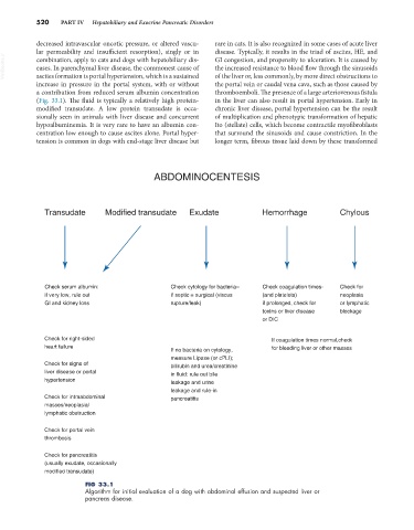
ABDOMINOCENTESIS
Transudate Modified transudate Exudate Hemorrhage Chylous
Check serum albumin: Check cytology for bacteria– Check coagulation times- Check for
if very low, rule out if septic = surgical (viscus (and platelets) neoplasia
GI and kidney loss rupture/leak) if prolonged, check for or lymphatic
toxins or liver disease blockage
or DIC
Check for right-sided If coagulation times normal,check
heart failure for bleeding liver or other masses
If no bacteria on cytology,
measure Lipase (or cPLI);
Check for signs of bilirubin and urea/creatinine
liver disease or portal in fluid: rule out bile
hypertension leakage and urine
leakage and rule-in
Check for intraabdominal pancreatitis
masses/neoplasia/
lymphatic obstruction
Check for portal vein
thrombosis
Check for pancreatitis
(usually exudate, occasionally
modified transudate)
FIG 33.1
Algorithm for initial evaluation of a dog with abdominal effusion and suspected liver or
pancreas disease.