
Page 553 - Small Animal Internal Medicine, 6th Edition
P. 553
CHAPTER 33 Clinical Manifestations of Hepatobiliary and Pancreatic Disease 525
hyperplasia, tend to result in smooth or slightly irregular,
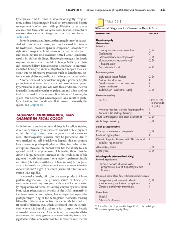
firm, diffuse hepatomegaly. Focal or asymmetrical hepatic TABLE 33.1
VetBooks.ir enlargement is often seen with proliferative or expansive Differential Diagnoses for Changes in Hepatic Size
diseases that form solid or cystic mass lesions. Examples of
diseases that cause a change in liver size are listed in
Table 33.1. DIAGNOSIS SPECIES
Smooth generalized hepatosplenomegaly may be associ- Hepatomegaly
ated with nonhepatic causes, such as increased intravascu- Generalized
lar hydrostatic pressure (passive congestion) secondary to Infiltration
right-sided congestive heart failure or pericardial disease. In Primary or metastatic neoplasia C, D
rare cases, hepatic vein occlusion (Budd-Chiari syndrome) Cholangitis C
results in similar findings. Hepatosplenomegaly in icteric Extramedullary hematopoiesis* C, D
Mononuclear-phagocytic cell
C, D
dogs or cats may be attributable to benign MPS hyperplasia hyperplasia*
and extramedullary hematopoiesis secondary to immune- Amyloidosis (rare) C, D
mediated hemolytic anemia. Hepatosplenomegaly may also
occur due to infiltrative processes such as lymphoma, sys- Passive congestion
temic mast cell disease, malignant histiocytosis, or leukemias. Right-sided heart failure C, D
Another cause of hepatosplenomegaly is primary hepatic Pericardial disease D
parenchymal disease with sustained intrahepatic portal Caudal vena cava obstruction D
hypertension. In dogs and cats with this syndrome, the liver Caval syndrome D
is usually firm and irregular on palpation, and often the liver Budd-Chiari syndrome (rare) C, D
itself is reduced in size as a result of fibrosis. However, the Hepatocyte swelling
spleen can be enlarged and congested as a result of portal Lipidosis C (moderate
hypertension. For conditions that involve primarily the to marked),
spleen, see Chapter 88. D (mild)
Hypercortisolism (steroid hepatopathy) D
Anticonvulsant drug therapy D
JAUNDICE, BILIRUBINURIA, AND Acute extrahepatic bile duct obstruction C, D
CHANGE IN FECAL COLOR
Acute hepatotoxicity C, D
By definition, jaundice in cats and dogs is the yellow staining Focal or asymmetric
of serum or tissues by an excessive amount of bile pigment Primary or metastatic neoplasia C, D
or bilirubin (Fig. 33.6); the terms jaundice and icterus are Nodular hyperplasia D
used interchangeably. Jaundice may be prehepatic, due to
very marked red cell breakdown; hepatic, due to primary Chronic hepatic disease with fibrosis and D
nodular regeneration
liver disease; or posthepatic, due to biliary tract obstruction
or rupture. Because the normal liver has the ability to take Abscess(es) (rare) C, D
up and excrete a large amount of bilirubin, there must be Cysts (rare) C, D
either a large, persistent increase in the production of bile Microhepatia (Generalized Only)
pigment (hyperbilirubinemia) or a major impairment in bile Reduced hepatic mass
excretion (cholestasis with hyperbilirubinemia) before jaun- Chronic hepatic disease with D
dice is detectable as yellow-stained tissues (serum bilirubin progressive loss of hepatocytes and
concentration ≥2 mg/dL) or serum (serum bilirubin concen- fibrosis
tration ≥1.5 mg/dL).
In normal animals, bilirubin is a waste product of heme Decreased portal blood flow with hepatocellular atrophy
protein degradation. The primary source of heme pro- Congenital portosystemic shunt C, D
teins is senescent erythrocytes, with a small contribution Intrahepatic portal vein hypoplasia D
by myoglobin and heme-containing enzyme systems in the Chronic portal vein thrombosis D
liver. After phagocytosis by cells of the MPS, primarily in Hypovolemia
the bone marrow and spleen, heme oxygenase opens the
protoporphyrin ring of the hemoglobin molecule, forming Shock? ?
biliverdin. Biliverdin reductase then converts biliverdin to Addison’s disease D
fat-soluble bilirubin IXa, which is released into the circula- C, Primarily cats; D, primarily dogs; C, D, cats and dogs.
tion, where it is bound to albumin for transport to hepatic *Concurrent splenomegaly likely.
sinusoidal membranes. After uptake, transhepatocellular
movement, and conjugation to various carbohydrates, con-
jugated bilirubin, now water-soluble, is excreted into the bile