
Page 555 - Small Animal Internal Medicine, 6th Edition
P. 555
CHAPTER 33 Clinical Manifestations of Hepatobiliary and Pancreatic Disease 527
Traumatic or pathologic biliary tract rupture allows Several nonhepatobiliary disorders impede bilirubin
leakage of bile into the peritoneal space and some absorption excretion by poorly understood means. Jaundice with evi-
VetBooks.ir of bile components. Depending on the underlying cause and dence of hepatocellular dysfunction but minimal histopath-
ologic changes in the liver has been described in septic
the time elapsed between biliary rupture and diagnosis, the
degree of jaundice may be mild to moderate. If biliary
by bacteria, such as endotoxin, are known to interfere with
rupture has occurred, the total bilirubin content of the human, feline, and canine patients. Certain products released
abdominal effusion is higher than that of serum. bile flow reversibly. As yet unexplained mild hyperbilirubi-
Reference ranges for serum total bilirubin concentrations nemia (≤2.5 mg/dL) may also be detected in approximately
in dogs and cats may vary among laboratories, but most 20% of hyperthyroid cats. Experimental investigations of
published resources agree that concentrations over 0.3 mg/ thyrotoxicosis in laboratory animals have demonstrated
dL in cats and 0.6 mg/dL in dogs are abnormal. When results increased production of bilirubin, which has been proposed
of laboratory tests are assessed, species differences in the to be associated with increased degradation of hepatic heme
formation and renal processing of bilirubin between cats and proteins. There is no histologic evidence of cholestasis at the
dogs must be taken into account. Canine renal tubules have light microscopic level in affected cats, and the hyperbiliru-
a low resorptive threshold for bilirubin. Dogs (males to a binemia resolves with return to euthyroidism. Guidelines for
greater extent than females) have the necessary renal enzyme initial evaluation of the icteric cat or dog are given in Fig.
systems to process bilirubin to a limited extent; therefore 33.9. Finally, lipemia is a common cause of pseudohyperbili-
bilirubinuria (up to 2+ to 3+ reaction by dipstick analysis) rubinemia in dogs as a result of lipid interference with the
may be a normal finding in canine urine specimens with a colorimetric laboratory methods.
specific gravity greater than 1.025. Cats do not have this Acholic feces result from the total absence of bile pigment
ability, and they have a ninefold higher tubular absorptive in the intestine (Fig. 33.10). Only a small amount of bile
capacity for bilirubin than dogs. Bilirubinuria in cats is asso- pigment is needed to be changed to stercobilin and yield
ciated with hyperbilirubinemia and is always pathologic a normal fecal color; therefore bile flow into the intestine
(Fig. 33.8). Because unconjugated and most conjugated bili- must be completely discontinued to result in acholic feces,
rubin is albumin-bound in the circulation, only a small and this is very rare in dogs and cats. In addition to appear-
amount of non–protein-bound conjugated bilirubin is ing pale from lack of stercobilin and other pigments, acholic
expected to appear in the urine in physiologic and patho- feces are pale because of steatorrhea resulting from the lack
logic states. In dogs with hepatobiliary disease, increasing of bile acids to facilitate fat absorption. Mechanical diseases
bilirubinuria often precedes the development of hyperbiliru- of the extrahepatic biliary tract (e.g., unremitting com-
binemia, and clinical jaundice and may be the first sign of plete EBDO, traumatic bile duct avulsion from the duode-
illness detected by owners. num) are the most common causes of acholic feces in cats
and dogs.
Feces also become pale as a result of exocrine pancreatic
insufficiency in dogs and cats, either as a result of pancreatic
acinar atrophy of end-stage chronic pancreatitis and marked
loss of pancreatic tissue mass. Fat maldigestion caused by a
lack of pancreatic lipase result in pale yellow, voluminous,
and smelly feces. Pancreatic enzyme lack can also be tran-
sient for example in an acute flare-up of chronic pancreatitis,
which may also cause biliary obstruction resulting in two
mechanisms for pale feces (see Fig. 33.10).
COAGULOPATHIES
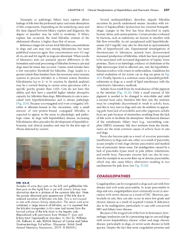
FIG 33.8 Coagulopathies can be recognized in dogs and cats with liver
Samples of urine (two pots on the left) and gallbladder bile disease and with acute pancreatitis. In acute pancreatitis in
(two pots on the right) from a cat with chronic biliary tract
obstruction due to a stricture of the common bile duct. Note dogs and cats, coagulopathies most commonly occur in asso-
the grossly abnormal pale appearance of the bile due to ciation with severe disease as a result of DIC. However, par-
reduced excretion of bilirubin into bile. This is not unusual ticularly in cats, they can also occur in more low grade and
in cats with chronic biliary obstruction. The serum and urine chronic disease as a result of acquired vitamin K deficiency
contained a large amount of bilirubin, so it is assumed that due to fat maldigestion, particularly in cats with concurrent
the bilirubin transporters in this case had moved from the IBD and biliary tract disease.
luminal surface of the bile ducts to the hepatic side. Because of the integral role of the liver in hemostasis, hem-
(Reproduced with permission from Watson P. Liver and
biliary tract: hepatocellular disorders. In: Hall EJ, Williams orrhagic tendencies can be a presenting sign in cats and dogs
DA, Kathrani A, eds. BSAVA Manual of Canine and Feline with severe hepatobiliary disease: either end-stage chronic
Gastroenterology 3rd edition. Gloucester: British Small disease, particularly in dogs, or severe acute disease in both
Animal Veterinary Association, 2019. © BSAVA.) species. Despite the fact that most coagulation proteins and