
Page 552 - Small Animal Internal Medicine, 6th Edition
P. 552
524 PART IV Hepatobiliary and Exocrine Pancreatic Disorders
recent study in dogs confirmed that animals with congenital status epilepticus, or coma will; prolonged severe HE by itself
PSS and symptomatic HE had higher serum C-reactive may lead to serious cerebral edema as a result of accumula-
VetBooks.ir protein concentrations than dogs with congenital PSS and tion of the osmolyte glutamine (from ammonia detoxifica-
tion) in astrocytes. In addition, the systemic effects of acute
no HE. C-reactive protein, an acute-phase protein, is a sensi-
tive nonspecific marker of inflammation in dogs, so this
and treated.
study adds support to the theory that inflammation may HE, particularly hypoglycemia, can be fatal if not recognized
trigger symptomatic HE in dogs with PSS. Furthermore, suc-
cessful ligation of congenital PSS resulted in the reduction
of inflammatory indicators in the blood as well as resolution CHANGE IN LIVER SIZE
of HE. In the author’s experience, it is often initially unde-
tected infections in the urinary tract, particularly pyelone- In normal cats and dogs, the liver is palpable just caudal to
phritis or cystitis, which trigger HE in susceptible dogs. the costal arch along the ventral body wall, but it may not
These may be acting in two ways: partly through production be palpable at all. Inability to palpate the liver, especially in
of inflammatory cytokines but also partly through absorp- dogs, does not automatically mean that the liver is small. In
tion of ammonia produced by urease-producing bacteria in lean cats, it is usually possible to palpate the diaphragmatic
the urinary tract. surface of the liver. In cats or dogs with pleural effusion or
other diseases that expand thoracic volume, the liver may
CLINICAL SIGNS be displaced caudally and give the false appearance of being
HE in humans is described as overt HE when there are enlarged.
obvious clinical signs and covert HE when signs are only Liver enlargement (hepatomegaly) is much more common
picked up on neuropyschometric testing. Such testing is not in cats than in dogs with liver disease. Dogs more often have
undertaken in dogs and cats, so only the more severe, overt a reduced liver size because of chronic hepatitis with fibrosis
HE is recognized. Subtle, nonspecific signs of HE in cats and but can show hepatomegaly particularly in association with
dogs that could be noted at any time and that represent tumors. The pattern of liver enlargement may be generalized
chronic or subclinical HE include anorexia, depression, or focal, depending on the cause. Infiltrative and congestive
weight loss, lethargy, nausea, fever, hypersalivation (particu- disease processes, or those that stimulate hepatocellular
larly in cats), intermittent vomiting, and diarrhea. Almost hypertrophy or mononuclear-phagocytic system (MPS)
any central nervous system (CNS) sign may be observed in
cats and dogs with HE, although typical signs tend to be
nonlocalizing, suggesting generalized brain involvement—
trembling, ataxia, hysteria, dementia, marked personality
change (usually toward aggressiveness), circling, head press-
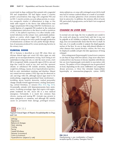
ing, cortical blindness, or seizures (Box 33.3; Fig. 33.5).
Occasionally, animals with hyperammonemia have asym-
metric, localizing neurologic signs that regress with appro-
priate treatment for HE. Acute HE is a true medical
emergency. Fortunately, it is much less common than
chronic, waxing and waning HE. Animals may present in
status epilepticus or comatose, and although HE initially
causes no permanent brain damage, prolonged seizures,
BOX 33.3
Typical Clinical Signs of Hepatic Encephalopathy in Dogs
and Cats
Lethargy
Depression
Behavioral changes
Head pressing
Circling
Pacing
Central blindness
Seizures (uncommon)
Coma (uncommon) FIG 33.5
Hypersalivation (especially cats) Head-pressing is one manifestation of hepatic
encephalopathy (Courtesy Georgina Harris).