
Page 549 - Small Animal Internal Medicine, 6th Edition
P. 549
CHAPTER 33 Clinical Manifestations of Hepatobiliary and Pancreatic Disease 521
stellate cells results in more irreversible sinusoidal obstruc- sodium delivery to the tubules and partly as a result of
tion. Thus the most common cause of portal hypertension increased release of renin-angiotensin-aldosterone (RAAS)
VetBooks.ir is chronic hepatitis progressing to cirrhosis in dogs (Fig. that results in increased sodium retention in the distal
tubules. This leads to an increase in circulating fluid volume,
33.2). It can also occur in association with hepatic neopla-
sia or in acute liver disease as a result of diffuse hepatic
reduces venous return because of increased pressure on the
swelling. In one study of acute liver disease, 41% of dogs which precipitates the formation of ascites, which in turn
had ascites. caudal vena cava and initiates a vicious cycle of renal sodium
The pathogenesis of the development of ascites in portal retention and ascites. This is the “over-fill” theory of ascites
hypertension is complex and has really been studied only in formation in liver disease. Therefore aldosterone antagonists
humans; it is assumed that the mechanisms of ascites are (e.g., spironolactone) are usually most effective in dogs with
similar in dogs. One way in which dogs differ from humans ascites secondary to portal hypertension, whereas loop
is that dogs do not develop the “spontaneous” infection of diuretics, such as furosemide used alone, can be ineffective
ascites of liver origin by extension of gut bacteria into the or even, in some cases, actually increase the volume of effu-
fluid that results in peritonitis, which is commonly reported sion by causing a further decrease in systemic blood pressure
in people. The presence of ascites is a poor prognostic indica- as a result of hemoconcentration and secondary increases in
tor in humans with chronic hepatitis, and the same appears RAAS activation.
to be true in dogs. Sodium retention by the kidneys is an Ascites will also occur as a result of venous congestion
important mechanism in the development of ascites in liver from disease of the major hepatic veins and/or distally (i.e.,
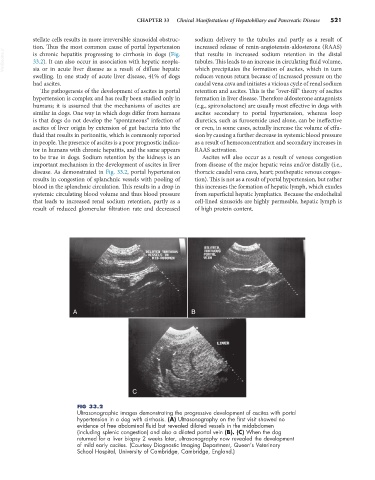
disease. As demonstrated in Fig. 33.2, portal hypertension thoracic caudal vena cava, heart; posthepatic venous conges-
results in congestion of splanchnic vessels with pooling of tion). This is not as a result of portal hypertension, but rather
blood in the splanchnic circulation. This results in a drop in this increases the formation of hepatic lymph, which exudes
systemic circulating blood volume and thus blood pressure from superficial hepatic lymphatics. Because the endothelial
that leads to increased renal sodium retention, partly as a cell-lined sinusoids are highly permeable, hepatic lymph is
result of reduced glomerular filtration rate and decreased of high protein content.
A B
C
FIG 33.2
Ultrasonographic images demonstrating the progressive development of ascites with portal
hypertension in a dog with cirrhosis. (A) Ultrasonography on the first visit showed no
evidence of free abdominal fluid but revealed dilated vessels in the midabdomen
(including splenic congestion) and also a dilated portal vein (B). (C) When the dog
returned for a liver biopsy 2 weeks later, ultrasonography now revealed the development
of mild early ascites. (Courtesy Diagnostic Imaging Department, Queen’s Veterinary
School Hospital, University of Cambridge, Cambridge, England.)