
Page 550 - Small Animal Internal Medicine, 6th Edition
P. 550
522 PART IV Hepatobiliary and Exocrine Pancreatic Disorders
PATHOGENESIS OF EXUDATES in dogs with cirrhosis and acquired PSS secondary to portal
In pancreatitis, the cause of abdominal fluid accumulation hypertension, in dogs and cats with acute liver failure, and
VetBooks.ir is usually increased vascular permeability due to peritonitis, in cats with hepatic lipidosis (which is in effect a reversible
acute liver failure). In a study of dogs with acute liver failure,
which in most cases results in serosanguinous sterile exu-
dates with high protein and cell contents, although modified
dogs suggest HE in about 7% of cases.
transudates and chylous effusions are occasionally reported. 57% showed signs of HE, and reports of chronic hepatitis in
Pancreatitis can also occasionally result in a more generalized The most important toxin implicated is ammonia (NH 3 ),
vasculitis and bicavitary effusions. Demonstrating a sterile which crosses the blood-brain barrier. The brain is very sen-
exudate on cytology should trigger investigations to differen- sitive to the toxic effects of NH 3 but does not possess a urea
tiate bile peritonitis, pancreatitis, or urinary tract leakage as cycle, so the NH 3 is detoxified by conversion to glutamine by
the causes, as detailed in Fig. 33.1. Urea, creatinine, bilirubin, astrocytes. Excessive NH 3 and thus glutamine buildup results
lipase, or cPLI concentrations can be measured in the fluid in osmotic stress of astrocytes with cell swelling and cerebral
and be very helpful diagnostically. Extravasation of bile from edema. Recent studies in dogs with congenital PSS have con-
a ruptured biliary tract elicits a strong inflammatory response firmed increases in arterial, venous, and CSF NH 3 when
and stimulates the transudation of lymph by serosal surfaces. compared with normal dogs. Sophisticated imaging tech-
In experimental animal models, the damaging component niques (single photon emission tomography) have also dem-
of bile has been identified as bile acids. Unlike with most onstrated reduced perfusion in the temporal lobes and
other causes of abdominal effusion associated with hepatobi- increased perfusion in the subcortical regions of dogs with
liary disease, there may be evidence of cranial abdominal or congenital PSS and HE, which are similar to the findings in
diffuse abdominal pain identified during physical examina- humans with acquired PSS and HE. Other toxins implicated
tion in cats and dogs with bile peritonitis. The fluid appears in HE historically such as mercaptans and aromatic amino
characteristically dark orange, yellow, or green and has a acids have little evidence in humans or animals, although
high bilirubin content on analysis, which is higher than the both manganese and neurosteroids have been implicated in
serum bilirubin concentration, and the predominant cell both species recently. Serum manganese concentrations are
type is the healthy neutrophil, except when the biliary tract high in dogs with congenital PSS, although the clinical rel-
is infected. Because normal bile is sterile, the initial phase of evance of this finding is unclear because ligation of con-
bile peritonitis is nonseptic, but unless treatment is initiated genital PSS in dogs resolves HE while hypermagnesemia
rapidly, secondary infection, usually with anaerobes of gut persists.
origin, may become life-threatening. Buildup of NH 3 in the systemic circulation occurs as a
Perivenular pyogranulomatous infiltrates in the visceral result of diversion of portal flow from the liver by the devel-
and parietal peritoneum of cats with the effusive form of FIP opment of PSS or as a result of a marked reduction in func-
increase vascular permeability and promote the exudation of tional hepatic mass. In most cases of acquired portosystemic
straw-colored, protein-rich fluid into the peritoneal space. shunting, there is a combination of vascular and functional
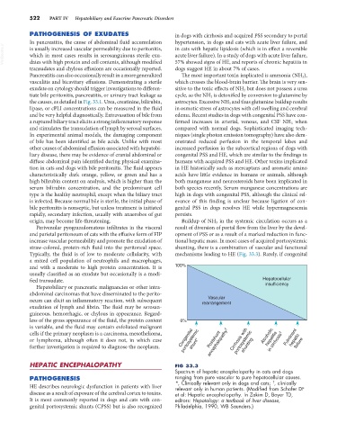
Typically, the fluid is of low to moderate cellularity, with mechanisms leading to HE (Fig. 33.3). Rarely, if congenital
a mixed cell population of neutrophils and macrophages,
and with a moderate to high protein concentration. It is 100%
usually classified as an exudate but occasionally is a modi-
fied transudate. Hepatocellular
Hepatobiliary or pancreatic malignancies or other intra- insufficiency
abdominal carcinomas that have disseminated to the perito-
Vascular
neum can elicit an inflammatory reaction, with subsequent rearrangement
exudation of lymph and fibrin. The fluid may be serosan-
guineous, hemorrhagic, or chylous in appearance. Regard-
less of the gross appearance of the fluid, the protein content 0%
is variable, and the fluid may contain exfoliated malignant
encephalopathy †
portosystemic
portosystemic
cells if the primary neoplasm is a carcinoma, mesothelioma, Alcoholic
in cirrhosis †
or lymphoma, although often it does not, in which case Congenital Postshunt Cirrhosis with hepatitis Fulminant hepatic failure
further investigation is required to diagnose the neoplasm. shunt* shunting
HEPATIC ENCEPHALOPATHY FIG 33.3
Spectrum of hepatic encephalopathy in cats and dogs
PATHOGENESIS ranging from pure vascular to pure hepatocellular causes.
†
HE describes neurologic dysfunction in patients with liver *, Clinically relevant only in dogs and cats; , clinically
relevant only in human patients. (Modified from Schafer DF
disease as a result of exposure of the cerebral cortex to toxins. et al: Hepatic encephalopathy. In Zakim D, Boyer TD,
It is most commonly reported in dogs and cats with con- editors: Hepatology: a textbook of liver disease,
genital portosystemic shunts (CPSS) but is also recognized Philadelphia, 1990, WB Saunders.)