
Page 79 - Small Animal Internal Medicine, 6th Edition
P. 79
CHAPTER 2 Diagnostic Tests for the Cardiovascular System 51
VetBooks.ir
A B
C
D E
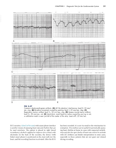
FIG 2.37
Common electrocardiogram artifacts. (A) 60 Hz electrical interference; lead III, 25 mm/
sec, dog. (B) Baseline movement caused by panting; lead II, 25 mm/sec, dog. (C)
Respiratory motion artifact; lead V 3, 50 mm/sec, dog. (D) Severe muscle tremor artifact;
lead V 3, 50 mm/sec, cat. (E) Intermittent, rapid baseline spikes caused by purring in cat;
a calibration mark is seen just left of the center of the strip. Lead aVF, 25 mm/sec.
ECG monitor, AliveCorVet.com) with smart phone interface has been recorded, it is sent via email to the veterinarian for
is another means of assessing heart rate and rhythm that can evaluation. This method can be useful for periodically assess-
be used anywhere. The patient is placed in right lateral ing heart rhythm at home in cases with suspected arrhyth-
recumbency, alcohol is applied to improve skin contact with mias and also for spot checks of heart rate control in animals
electrodes on the back of the recorder, the device (with with AF. Artifacts can hinder interpretation in some cases,
linked smart phone) is positioned on the chest wall over the especially in those patients that are not quiet and relaxed
heart, and the recording function is activated. After the ECG during recording.