
Page 75 - Small Animal Internal Medicine, 6th Edition
P. 75
CHAPTER 2 Diagnostic Tests for the Cardiovascular System 47
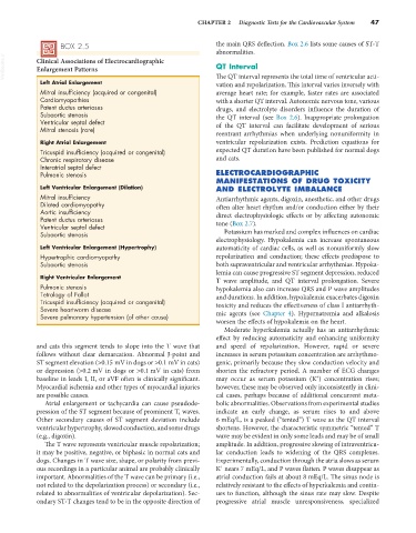
BOX 2.5 the main QRS deflection. Box 2.6 lists some causes of ST-T
abnormalities.
VetBooks.ir Clinical Associations of Electrocardiographic QT Interval
Enlargement Patterns
Left Atrial Enlargement The QT interval represents the total time of ventricular acti-
vation and repolarization. This interval varies inversely with
Mitral insufficiency (acquired or congenital) average heart rate; for example, faster rates are associated
Cardiomyopathies with a shorter QT interval. Autonomic nervous tone, various
Patent ductus arteriosus drugs, and electrolyte disorders influence the duration of
Subaortic stenosis the QT interval (see Box 2.6). Inappropriate prolongation
Ventricular septal defect of the QT interval can facilitate development of serious
Mitral stenosis (rare)
reentrant arrhythmias when underlying nonuniformity in
Right Atrial Enlargement ventricular repolarization exists. Prediction equations for
Tricuspid insufficiency (acquired or congenital) expected QT duration have been published for normal dogs
Chronic respiratory disease and cats.
Interatrial septal defect
Pulmonic stenosis ELECTROCARDIOGRAPHIC
MANIFESTATIONS OF DRUG TOXICITY
Left Ventricular Enlargement (Dilation) AND ELECTROLYTE IMBALANCE
Mitral insufficiency Antiarrhythmic agents, digoxin, anesthetic, and other drugs
Dilated cardiomyopathy often alter heart rhythm and/or conduction either by their
Aortic insufficiency direct electrophysiologic effects or by affecting autonomic
Patent ductus arteriosus
Ventricular septal defect tone (Box 2.7).
Subaortic stenosis Potassium has marked and complex influences on cardiac
electrophysiology. Hypokalemia can increase spontaneous
Left Ventricular Enlargement (Hypertrophy) automaticity of cardiac cells, as well as nonuniformly slow
Hypertrophic cardiomyopathy repolarization and conduction; these effects predispose to
Subaortic stenosis both supraventricular and ventricular arrhythmias. Hypoka-
lemia can cause progressive ST segment depression, reduced
Right Ventricular Enlargement T wave amplitude, and QT interval prolongation. Severe
Pulmonic stenosis hypokalemia also can increase QRS and P wave amplitudes
Tetralogy of Fallot and durations. In addition, hypokalemia exacerbates digoxin
Tricuspid insufficiency (acquired or congenital) toxicity and reduces the effectiveness of class I antiarrhyth-
Severe heartworm disease
Severe pulmonary hypertension (of other cause) mic agents (see Chapter 4). Hypernatremia and alkalosis
worsen the effects of hypokalemia on the heart.
Moderate hyperkalemia actually has an antiarrhythmic
effect by reducing automaticity and enhancing uniformity
and cats this segment tends to slope into the T wave that and speed of repolarization. However, rapid or severe
follows without clear demarcation. Abnormal J-point and increases in serum potassium concentration are arrhythmo-
ST segment elevation (>0.15 mV in dogs or >0.1 mV in cats) genic, primarily because they slow conduction velocity and
or depression (>0.2 mV in dogs or >0.1 mV in cats) from shorten the refractory period. A number of ECG changes
+
baseline in leads I, II, or aVF often is clinically significant. may occur as serum potassium (K ) concentration rises;
Myocardial ischemia and other types of myocardial injuries however, these may be observed only inconsistently in clini-
are possible causes. cal cases, perhaps because of additional concurrent meta-
Atrial enlargement or tachycardia can cause pseudode- bolic abnormalities. Observations from experimental studies
pression of the ST segment because of prominent T a waves. indicate an early change, as serum rises to and above
Other secondary causes of ST segment deviation include 6 mEq/L, is a peaked (“tented”) T wave as the QT interval
ventricular hypertrophy, slowed conduction, and some drugs shortens. However, the characteristic symmetric “tented” T
(e.g., digoxin). wave may be evident in only some leads and may be of small
The T wave represents ventricular muscle repolarization; amplitude. In addition, progressive slowing of intraventricu-
it may be positive, negative, or biphasic in normal cats and lar conduction leads to widening of the QRS complexes.
dogs. Changes in T wave size, shape, or polarity from previ- Experimentally, conduction through the atria slows as serum
+
ous recordings in a particular animal are probably clinically K nears 7 mEq/L, and P waves flatten. P waves disappear as
important. Abnormalities of the T wave can be primary (i.e., atrial conduction fails at about 8 mEq/L. The sinus node is
not related to the depolarization process) or secondary (i.e., relatively resistant to the effects of hyperkalemia and contin-
related to abnormalities of ventricular depolarization). Sec- ues to function, although the sinus rate may slow. Despite
ondary ST-T changes tend to be in the opposite direction of progressive atrial muscle unresponsiveness, specialized