
Page 71 - Small Animal Internal Medicine, 6th Edition
P. 71
CHAPTER 2 Diagnostic Tests for the Cardiovascular System 43
VetBooks.ir
A
B
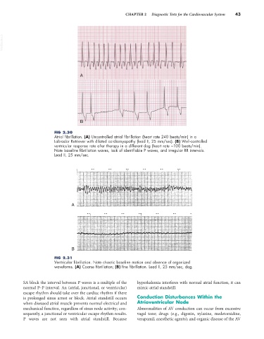
FIG 2.30
Atrial fibrillation. (A) Uncontrolled atrial fibrillation (heart rate 240 beats/min) in a
Labrador Retriever with dilated cardiomyopathy (lead II, 25 mm/sec). (B) Well-controlled
ventricular response rate after therapy in a different dog (heart rate ~100 beats/min).
Note baseline fibrillation waves, lack of identifiable P waves, and irregular RR intervals.
Lead II, 25 mm/sec.
A
B
FIG 2.31
Ventricular fibrillation. Note chaotic baseline motion and absence of organized
waveforms. (A) Coarse fibrillation; (B) fine fibrillation. Lead II, 25 mm/sec, dog.
SA block the interval between P waves is a multiple of the hyperkalemia interferes with normal atrial function, it can
normal P–P interval. An (atrial, junctional, or ventricular) mimic atrial standstill.
escape rhythm should take over the cardiac rhythm if there
is prolonged sinus arrest or block. Atrial standstill occurs Conduction Disturbances Within the
when diseased atrial muscle prevents normal electrical and Atrioventricular Node
mechanical function, regardless of sinus node activity; con- Abnormalities of AV conduction can occur from excessive
sequently, a junctional or ventricular escape rhythm results. vagal tone; drugs (e.g., digoxin, xylazine, medetomidine,
P waves are not seen with atrial standstill. Because verapamil, anesthetic agents); and organic disease of the AV