
Page 68 - Small Animal Internal Medicine, 6th Edition
P. 68
40 PART I Cardiovascular System Disorders
VetBooks.ir
A
B
C
D
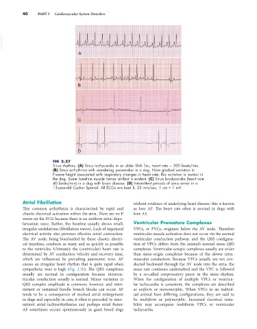
FIG 2.27
Sinus rhythms. (A) Sinus tachycardia in an older Shih Tzu; heart rate ~ 200 beats/min.
(B) Sinus arrhythmia with wandering pacemaker in a dog. Note gradual variation in
P-wave height associated with respiratory changes in heart rate; this variation is normal in
the dog. Some baseline muscle tremor artifact is evident. (C) Sinus bradycardia (heart rate
40 beats/min) in a dog with brain disease. (D) Intermittent periods of sinus arrest in a
13-year-old Cocker Spaniel. All ECGs are lead II, 25 mm/sec, 1 cm = 1 mV.
Atrial Fibrillation without evidence of underlying heart disease; this is known
This common arrhythmia is characterized by rapid and as lone AF. The heart rate often is normal in dogs with
chaotic electrical activation within the atria. There are no P lone AF.
waves on the ECG because there is no uniform atrial depo-
larization wave. Rather, the baseline usually shows small, Ventricular Premature Complexes
irregular undulations (fibrillation waves). Lack of organized VPCs, or PVCs, originate below the AV node. Therefore
electrical activity also prevents effective atrial contraction. ventricular muscle activation does not occur via the normal
The AV node, being bombarded by these chaotic electri- ventricular conduction pathway, and the QRS configura-
cal impulses, conducts as many and as quickly as possible tion of VPCs differs from the animal’s normal sinus QRS
to the ventricles. Ultimately the (ventricular) heart rate is complexes. Ventricular ectopic complexes usually are wider
determined by AV conduction velocity and recovery time, than sinus-origin complexes because of the slower intra-
which are influenced by prevailing autonomic tone. AF muscular conduction. Because VPCs usually are not con-
causes an irregular heart rhythm that is quite rapid when ducted backward through the AV node into the atria, the
sympathetic tone is high (Fig. 2.30). The QRS complexes sinus rate continues undisturbed and the VPC is followed
usually are normal in configuration because intraven- by a so-called compensatory pause in the sinus rhythm.
tricular conduction usually is normal. Minor variation in When the configuration of multiple VPCs or ventricu-
QRS complex amplitude is common, however, and inter- lar tachycardia is consistent, the complexes are described
mittent or sustained bundle branch blocks can occur. AF as uniform or monomorphic. When VPCs in an individ-
tends to be a consequence of marked atrial enlargement ual animal have differing configurations, they are said to
in dogs and especially in cats; it often is preceded by inter- be multiform or polymorphic. Increased electrical insta-
mittent atrial tachyarrhythmias and perhaps atrial flutter. bility may accompany multiform VPCs or ventricular
AF sometimes occurs spontaneously in giant breed dogs tachycardia.