
Page 66 - Small Animal Internal Medicine, 6th Edition
P. 66
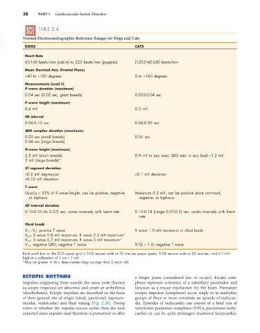
38     PART I   Cardiovascular System Disorders


                   TABLE 2.4
  VetBooks.ir  Normal Electrocardiographic Reference Ranges for Dogs and Cats

             DOGS
                                                                 CATS
             Heart Rate
             60-160 beats/min (adults) to 220 beats/min (puppies)  (120-)140-240 beats/min
             Mean Electrical Axis (Frontal Plane)
             +40 to +100 degrees                                 0 to +160 degrees
             Measurements (Lead II)
             P-wave duration (maximum)
             0.04 sec (0.05 sec, giant breeds)                   0.035-0.04 sec

             P-wave height (maximum)
             0.4 mV                                              0.2 mV

             PR interval
             0.06-0.13 sec                                       0.05-0.09 sec

             QRS complex duration (maximum)
             0.05 sec (small breeds)                             0.04 sec
             0.06 sec (large breeds)
             R-wave height (maximum)
             2.5 mV (small breeds)                               0.9 mV in any lead; QRS total in any lead <1.2 mV
             3 mV (large breeds)*

             ST segment deviation
             <0.2 mV depression                                  <0.1 mV deviation
             <0.15 mV elevation
             T wave
             Usually < 25% of R wave height; can be positive, negative,   Maximum 0.3 mV; can be positive (most common),
               or biphasic                                        negative, or biphasic

             QT interval duration
             0.15-0.25 (to 0.27) sec; varies inversely with heart rate  0.12-0.18 (range 0.07-0.2) sec; varies inversely with heart
                                                                  rate
             Chest Leads
             V 1 ; rV 2 : positive T wave                        R wave 1.0 mV maximum in chest leads
             V 2-3 : S wave 0.8 mV maximum; R wave 2.5 mV maximum*
             V 4-6 : S wave 0.7 mV maximum; R wave 3 mV maximum*
             V 10: negative QRS; negative T wave                 R/Q < 1.0; negative T wave

            Each small box on the ECG paper grid is 0.02 second wide at 50 mm/sec paper speed, 0.04 second wide at 25 mm/sec, and 0.1 mV
            high at a calibration of 1 cm = 1 mV.
            *May be greater in thin, deep-chested dogs younger than 2 years old.


            ECTOPIC RHYTHMS                                      a longer pause (considered late or  escape). Escape com-
            Impulses originating from outside the sinus node (known   plexes represent activation of a subsidiary pacemaker and
            as ectopic impulses) are abnormal and create an arrhythmia   function as a rescue mechanism for the heart. Premature
            (dysrhythmia). Ectopic impulses are described on the basis   ectopic impulses (complexes) occur singly or in multiples;
            of their general site of origin (atrial, junctional, supraven-  groups of three or more constitute an episode of tachycar-
            tricular, ventricular) and their timing (Fig. 2.28).  Timing   dia. Episodes of tachycardia can consist of a brief run of
            refers to whether the impulse occurs earlier than the next   ventricular premature complexes (VPCs; paroxysmal tachy-
            expected sinus impulse (and therefore is premature) or after   cardia) or can be quite prolonged (sustained tachycardia).