
Page 64 - Small Animal Internal Medicine, 6th Edition
P. 64
36 PART I Cardiovascular System Disorders
–90° –90°
VetBooks.ir –120° –60° –120° –60°
aVR aVL aVR
–30° –150° –30° aVL
–150°
RIGHT RIGHT
LEFT LEFT
±180° 0° ±180° 0°
I I
+150° +30° +150° +30°
CAUDAL CAUDAL
+120° +60° +120° +60°
III +90° II III +90° II
A aVF B aVF
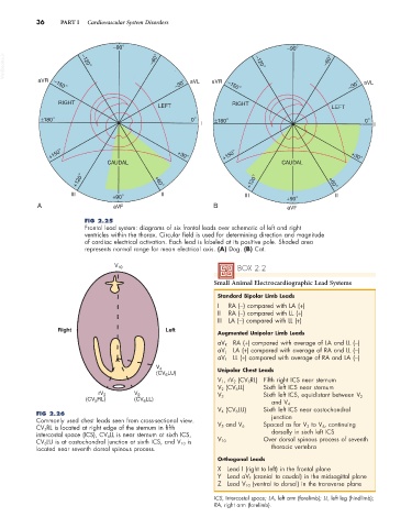
FIG 2.25
Frontal lead system: diagrams of six frontal leads over schematic of left and right
ventricles within the thorax. Circular field is used for determining direction and magnitude
of cardiac electrical activation. Each lead is labeled at its positive pole. Shaded area
represents normal range for mean electrical axis. (A) Dog. (B) Cat.
V 10 BOX 2.2
Small Animal Electrocardiographic Lead Systems
Standard Bipolar Limb Leads
I RA (−) compared with LA (+)
II RA (−) compared with LL (+)
III LA (−) compared with LL (+)
Right Left
Augmented Unipolar Limb Leads
aV R RA (+) compared with average of LA and LL (−)
aV L LA (+) compared with average of RA and LL (−)
aV F LL (+) compared with average of RA and LA (−)
V 4
(CV LU) Unipolar Chest Leads
6
V 1 , rV 2 (CV 5 RL) Fifth right ICS near sternum
V 2 (CV 6 LL) Sixth left ICS near sternum
rV 2 V 2 V 3 Sixth left ICS, equidistant between V 2
(CV RL) (CV LL)
6
5
and V 4
V 4 (CV 6 LU) Sixth left ICS near costochondral
FIG 2.26 junction
Commonly used chest leads seen from cross-sectional view. V 5 and V 6
CV 5RL is located at right edge of the sternum in fifth Spaced as for V 3 to V 4 , continuing
intercostal space (ICS), CV 6LL is near sternum at sixth ICS, dorsally in sixth left ICS
CV 6LU is at costochondral junction at sixth ICS, and V 10 is V 10 Over dorsal spinous process of seventh
located near seventh dorsal spinous process. thoracic vertebra
Orthogonal Leads
X Lead I (right to left) in the frontal plane
Y Lead aV F (cranial to caudal) in the midsagittal plane
Z Lead V 10 (ventral to dorsal) in the transverse plane
ICS, Intercostal space; LA, left arm (forelimb); LL, left leg (hindlimb);
RA, right arm (forelimb).