
Page 61 - Small Animal Internal Medicine, 6th Edition
P. 61
CHAPTER 2 Diagnostic Tests for the Cardiovascular System 33
VetBooks.ir
A B
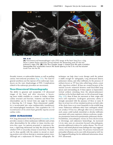
FIG 2.21
(A) Two-dimensional transesophageal echo (TEE) image at the heart base from a dog
shows a patent ductus arteriosus (arrow) between the descending aorta (D Ao) and
pulmonary artery (PA). (B) Color flow Doppler image in diastole from the same orientation
demonstrates flow acceleration toward the ductal opening in the D Ao and the turbulent
ductal flow into the PA.
thrombi, tumors, or endocarditis lesions, as well as guiding technique can help direct acute therapy until the patient
cardiac interventional procedures (Fig. 2.21). The need for is stable enough for radiography. Lung ultrasound detects
general anesthesia and the expense of the endoscopic trans- pulmonary edema, and other infiltrates, by the presence of
ducers are the main disadvantages of TEE. Complications artifacts known as “B” lines (also called lung rockets, comet
related to the endoscopy procedure are uncommon. tails, ring-down artifact). B lines are created because of the
marked acoustic mismatch between small fluid-filled lung
Three-Dimensional Echocardiography spaces and surrounding air. B lines appear as hyperechoic
The ability to generate and manipulate 3-D ultrasound vertical artifacts stretching from the pleural-pulmonary
images of the heart and other structures is becom- interface to the furthest depth seen on the ultrasound image
ing more widely available as a means to further evaluate (Fig. 2.22); these B lines are narrowest at their origin and
cardiac structure and function. Anatomic and blood flow move with respiration. Cardiogenic pulmonary edema is
abnormalities can be viewed from any angle by rotating strongly associated with the presence of three or more B
or bisecting the 3-D images. Three-dimensional capabil- lines in at least two of four standard positions on both sides
ity is incorporated into some multimodality transthoracic of the chest; these occur most frequently in the middle lung
and transesophageal transducers. Data acquisition for 3-D zones. Pulmonary infiltrates other than cardiogenic edema
reconstruction of the entire heart generally requires several can produce B lines too, although often not as extensively;
cardiac cycles. such causes could include interstitial or alveolar infiltrates
associated with acute respiratory distress syndrome, neopla-
LUNG ULTRASOUND sia, pneumonia, heartworm pneumonitis, pulmonary throm-
POC lung ultrasound (Vet BLUE protocol; Lisciandro, 2014) boembolism, noncardiogenic edema (as from electrocution
provides a means to detect pulmonary infiltrates and certain or drowning), and pulmonary hemorrhage. An increased
other abnormalities by the artifact or tissue interface pat- LA:Ao ratio, seen on concurrent focused echocardiogram,
terns they cause. For patients presented in respiratory dis- provides additional evidence for cardiac disease. The clinical
tress, a rapid lung ultrasound can help the clinician decide history and initial physical exam findings also may suggest a
whether CHF or noncardiac disease is most likely. The exam cardiac versus noncardiac cause. The presence of pleural and
can be done quickly with the patient in sternal or stand- pericardial effusions can be seen with ultrasound as well. See
ing position and without shaving to minimize patient stress. the Suggested Readings list for additional information about
Although not a replacement for thoracic radiographs, the POC ultrasound.