
Page 60 - Small Animal Internal Medicine, 6th Edition
P. 60
32 PART I Cardiovascular System Disorders
produces multiple velocities and directions of flow in an area mean myocardial velocities from different regions. Other
resulting in a mixing of color; this display can be enhanced techniques used to assess regional myocardial function and
VetBooks.ir using a variance map, which adds shades of yellow or green synchrony can be derived from DTI methods, including
myocardial velocity gradients, myocardial strain, and strain
to the red/blue display (Fig. 2.19).
The severity of valve regurgitation is estimated subjec-
tively by the size and shape of the regurgitant jet during CF rate.
Myocardial strain and strain rate indices can be helpful
imaging. Although technical and hemodynamic factors in assessing subclinical myocardial wall motion abnormali-
confound the accuracy of such assessment, wide and long ties and ventricular dyssynchrony. Strain is a measure of
regurgitant jets generally are associated with more severe myocardial deformation or percent change from its original
regurgitation than jets that are narrow at their point of dimension. Strain rate describes the temporal rate of defor-
origin. Other methods for quantifying valve regurgitation mation. A significant limitation of Doppler-based techniques
have been described as well. Maximum regurgitant jet veloc- is their angle dependence, complicated by cardiac transla-
ity is not a good indicator of severity, especially with mitral tional motion. A “speckle tracking” modality, based on 2-D
regurgitation. Changes in chamber size (i.e., eccentric hyper- echocardiography rather than DTI, is often used now as a
trophy and remodeling) provide a better indication of sever- potentially more accurate way to assess regional myocardial
ity with chronic regurgitation. motion, strain, and strain rate. This modality relies on track-
ing the motion of gray scale “speckles” within the myocar-
OTHER ECHOCARDIOGRAPHIC dium as they move throughout the cardiac cycle. More
MODALITIES information can be found in the Suggested Readings.
Doppler Tissue Imaging and 2-D
Speckle Tracking Transesophageal Echocardiography
Doppler tissue imaging (DTI) is a modality used to assess Transesophageal echocardiography (TEE) employs transduc-
the motion of tissue, rather than blood cells, by altering the ers mounted on a flexible, steerable endoscope tip to image
signal processing and filtering of returning echoes. Myocar- cardiac structures through the esophageal wall. TEE can
dial velocity patterns can be assessed with CF and PW spec- provide clearer images of some cardiac structures (especially
tral DTI techniques. Spectral DTI provides greater temporal those at or above the AV junction) compared with transtho-
resolution and quantifies velocity of myocardial motion at racic echocardiography because chest wall and lung interfer-
specific locations, such as the lateral or septal aspects of ence is avoided. This technique can be particularly useful
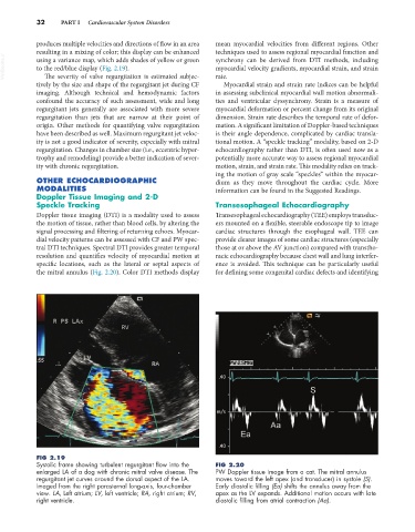
the mitral annulus (Fig. 2.20). Color DTI methods display for defining some congenital cardiac defects and identifying
FIG 2.19
Systolic frame showing turbulent regurgitant flow into the FIG 2.20
enlarged LA of a dog with chronic mitral valve disease. The PW Doppler tissue image from a cat. The mitral annulus
regurgitant jet curves around the dorsal aspect of the LA. moves toward the left apex (and transducer) in systole (S).
Imaged from the right parasternal long-axis, four-chamber Early diastolic filling (Ea) shifts the annulus away from the
view. LA, Left atrium; LV, left ventricle; RA, right atrium; RV, apex as the LV expands. Additional motion occurs with late
right ventricle. diastolic filling from atrial contraction (Aa).