
Page 69 - Small Animal Internal Medicine, 6th Edition
P. 69
CHAPTER 2 Diagnostic Tests for the Cardiovascular System 41
Ectopic complexes - origin represents a melding of the normal QRS configuration and
Normal sinus that of the VPC (see Fig. 2.29, F). Fusion complexes often
VetBooks.ir are observed at the onset or end of a paroxysm (run) of
ventricular tachycardia; they are preceded by a P wave and
Supraventricular (atrial or junctional) shortened PR interval. Identification of P waves (whether
conducted or not) or fusion complexes helps in differentiat-
ing ventricular tachycardia from SVT with abnormal (aber-
rant) intraventricular conduction.
Polymorphic ventricular tachycardia is characterized by
Ventricular
QRS complexes that vary in size, polarity, and often rate;
sometimes the QRS configuration appears as if it were rotat-
ing around the isoelectric baseline. Torsades de pointes is a
A specific form of polymorphic ventricular tachycardia associ-
ated with Q-T interval prolongation.
Ectopic complexes - timing
Accelerated Idioventricular Rhythm
Premature (early) Also called accelerated ventricular rhythm or idioventricular
Supraventricular (junctional, atrial)
tachycardia, an accelerated idioventricular rhythm originates
within the ventricles and has a rate of about 60 to 100 beats/
min in the dog (perhaps somewhat faster in the cat). Because
Ventricular the rate is slower than true ventricular tachycardia, it usually
is a less serious rhythm disturbance. An accelerated ven-
tricular rhythm can appear intermittently during sinus
arrhythmia, as the sinus rate decreases; then the idioven-
Escape (late) tricular rhythm usually is suppressed as the sinus rate
increases. This rhythm is common in dogs recovering from
motor vehicle trauma; it also occurs in many dogs with
B severe intra-abdominal or systemic disease. Often this
rhythm disturbance has no deleterious effects, although it
FIG 2.28 could progress to ventricular tachycardia, especially in clini-
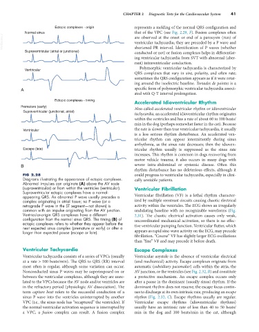
Diagrams illustrating the appearance of ectopic complexes. cally unstable patients.
Abnormal impulses can originate (A) above the AV node
(supraventricular) or from within the ventricles (ventricular). Ventricular Fibrillation
Supraventricular ectopic complexes have a normal-
appearing QRS. An abnormal P wave usually precedes a Ventricular fibrillation (VF) is a lethal rhythm character-
complex originating in atrial tissue; no P wave (or a ized by multiple reentrant circuits causing chaotic electrical
retrograde P wave in the ST segment—not shown) is activity within the ventricles. The ECG shows an irregularly
common with an impulse originating from the AV junction. undulating baseline with no recognizable waveforms (Fig.
Ventricular-origin QRS complexes have a different 2.31). The chaotic electrical activation causes only weak,
configuration from the normal sinus QRS. The timing (B) of uncoordinated mechanical activation, so there is no effec-
ectopic complexes refers to whether they appear before the tive ventricular pumping function. Ventricular flutter, which
next expected sinus complex (premature or early) or after a
longer than expected pause (escape or late). appears as rapid sine-wave activity on the ECG, may precede
fibrillation. “Course” VF has slightly larger ECG oscillations
than “fine” VF and may precede it before death.
Ventricular Tachycardia Escape Complexes
Ventricular tachycardia consists of a series of VPCs (usually Ventricular asystole is the absence of ventricular electrical
at a rate > 100 beats/min). The QRS to QRS (RR) interval (and mechanical) activity. Escape complexes originate from
most often is regular, although some variation can occur. automatic (subsidiary pacemaker) cells within the atria, the
Nonconducted sinus P waves may be superimposed on or AV junction, or the ventricles (see Fig. 2.32, B) and constitute
between the ventricular complexes, although they are unre- a protective mechanism. An escape complex occurs only
lated to the VPCs because the AV node and/or ventricles are after a pause in the dominant (usually sinus) rhythm. If the
in the refractory period (physiologic AV dissociation). The dominant rhythm does not resume, the escape focus contin-
term capture beat refers to the successful conduction of a ues to discharge at its own intrinsic rate, producing an escape
sinus P wave into the ventricles uninterrupted by another rhythm (Fig. 2.32, C). Escape rhythms usually are regular.
VPC (i.e., the sinus node has “recaptured” the ventricles). If Ventricular escape rhythms (idioventricular rhythms)
the normal ventricular activation sequence is interrupted by usually have an intrinsic rate of less than 40 to 50 beats/
a VPC, a fusion complex can result. A fusion complex min in the dog and 100 beats/min in the cat, although