
Page 70 - Small Animal Internal Medicine, 6th Edition
P. 70
42 PART I Cardiovascular System Disorders
VetBooks.ir
A B
C D
E
F
H
G
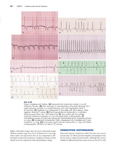
FIG 2.29
Ectopic complexes and rhythms. (A) Supraventricular premature complex in an old
Doberman Pinscher. (B) Short paroxysm of supraventricular tachycardia (bracket) with 1
single premature complex in a mixed-breed dog with mitral regurgitation (lead II,
25 mm/sec). (C) Sustained atrial tachycardia in Irish Setter with mitral stenosis. Note
negative, abnormal P waves. (D) Rapid supraventricular tachycardia in a Labrador
Retriever puppy (heart rate ~360 beats/min). (E) Sinus rhythm with two isolated
ventricular premature complexes in a cat with hypertrophic cardiomyopathy. (F)
Intermittent paroxysms of ventricular tachycardia demonstrating fusion complexes (arrows)
in a dog. (G) Accelerated idioventricular rhythm interspersed with a background of sinus
arrhythmia in a dog. (H) Paroxysmal, rapid ventricular tachycardia in a dog with dilated
cardiomyopathy. All ECGs are lead II, 25 mm/sec.
higher ventricular escape rates can occur. Junctional escape CONDUCTION DISTURBANCES
rhythms usually range from 40 to 60 beats/min in the dog, Abnormal impulse conduction within the atria can occur at
with a faster rate expected in the cat. It is important to dif- several sites. SA block prevents impulse transmission from
ferentiate escape from premature complexes. Escape activity the SA node to adjacent atrial muscle. Although this cannot
should never be suppressed with antiarrhythmic drugs. reliably be differentiated from sinus arrest on the ECG, with