
Page 74 - Small Animal Internal Medicine, 6th Edition
P. 74
46 PART I Cardiovascular System Disorders
A right-axis deviation and an S wave in lead I are strong alters QRS configuration. Electrical activation in regions of
criteria for RV enlargement (or RBBB). Other ECG changes ventricular muscle served by the affected bundle branch
VetBooks.ir usually can be found as well. Three or more of the criteria occurs late and progresses slowly. This widens the QRS
complex (especially the terminal portion of it) and shifts the
listed in Box 2.4 generally are present when RV enlargement
exists. RV enlargement (dilation or hypertrophy) is likely to
be pronounced if it is evident on the ECG because LV activa- QRS (and MEA) orientation toward the area of delayed acti-
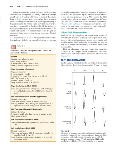
vation. Box 2.4 and Fig. 2.35 summarize ECG patterns asso-
tion forces are normally so dominant. LV dilation and eccen- ciated with ventricular enlargement or conduction delay.
tric hypertrophy typically increase R-wave amplitude in the Box 2.5 lists common clinical associations.
caudal leads (II and aVF) and sometimes widen the QRS. LV
concentric hypertrophy inconsistently produces a left-axis Other QRS Abnormalities
deviation. Small-voltage QRS complexes sometimes occur. Causes of
Conduction block in any of the major ventricular conduc- reduced QRS amplitude include pleural or pericardial effu-
tion pathways disturbs the normal activation process and sions, obesity, intrathoracic mass lesions, hypovolemia, and
hypothyroidism. Small complexes occasionally are seen in
dogs with dilated cardiomyopathy or without identifiable
BOX 2.4 abnormalities.
Electrical alternans is an every-other-beat recurring
Ventricular Chamber Enlargement and Conduction alteration in QRS complex size or configuration. This most
Abnormality Patterns
often is seen with large-volume pericardial effusions (see
Normal Chapter 9).
Normal mean electrical axis ST-T ABNORMALITIES
No S wave in lead I
R wave taller in lead II than in lead I The ST segment extends from the end of the QRS complex
Lead V 2 R wave larger than S wave (also called the J-point) to the onset of the T wave. In dogs
Right Ventricular Enlargement I II/aVF V V
Right-axis deviation 3 10
S wave present in lead I
S wave in V 2-3 larger than R wave or > 0.8 mV
Q-S (“W” shape) in V 10
Positive T wave in lead V 10
Deep S wave in leads II, III, and aV F Normal
Right Bundle Branch Block (RBBB)
Same as right ventricular enlargement, with prolonged
terminal (latter) portion of the QRS (wide, sloppy S
RVE
wave) (RBBB)
Left Ventricular Dilation (Eccentric Hypertrophy)
Normal frontal axis
Taller than normal R wave in leads II, aV F , V 2-3
Possibly widened QRS; slurring and displacement of ST
segment and T-wave enlargement may also occur
LV dilation
Left Ventricular (Concentric) Hypertrophy (LPFB)
Left-axis deviation
R wave in lead I taller than R wave in leads II or aV F
No S wave in lead I
Left Anterior Fascicular Block (LAFB)
Same as left ventricular hypertrophy, possibly with wider LV hypertrophy
(LAFB)
QRS
Left Bundle Branch Block (LBBB)
Normal frontal axis FIG 2.35
Very wide and sloppy QRS; R waves may be taller than Schematic of common ventricular enlargement patterns and
conduction abnormalities. Electrocardiogram leads are listed
normal in leads II, aV F, V 2-3
Small Q wave may be present in leads II, III, and aV F across top. LAFB, Left anterior fascicular block; LPFB, left
(incomplete LBBB) posterior fascicular block; LV, left ventricular; RBBB, right
bundle branch block; RVE, right ventricular enlargement.