
Page 77 - Small Animal Internal Medicine, 6th Edition
P. 77
CHAPTER 2 Diagnostic Tests for the Cardiovascular System 49
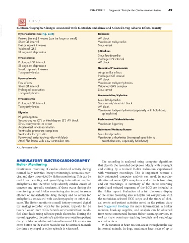
BOX 2.7
VetBooks.ir Electrocardiographic Changes Associated With Electrolyte Imbalance and Selected Drug Adverse Effects/Toxicity
Hyperkalemia (See Fig. 2.36)
Lidocaine
Peaked (tented) T waves (can be large or small) AV block
Short QT interval Ventricular tachycardia
Flat or absent P waves Sinus arrest
Widened QRS
ST segment depression β-Blockers
Sinus bradycardia
Hypokalemia Prolonged PR interval
Prolonged QT interval AV block
ST segment depression
Small, biphasic T waves Quinidine/Procainamide
Tachyarrhythmias Atropine-like effects
Prolonged QT interval
Hypercalcemia AV block
Few effects Ventricular tachyarrhythmias
Short QT interval Widened QRS complex
Prolonged conduction Sinus arrest
Tachyarrhythmias
Medetomidine/Xylazine
Hypocalcemia Sinus bradycardia
Prolonged QT interval Sinus arrest/sinoatrial block
Tachyarrhythmias AV block
Ventricular tachyarrhythmias (especially with halothane,
Digoxin epinephrine)
PR prolongation
Second-degree (2°) or third-degree (3°) AV block Barbiturates/Thiobarbiturates
Sinus bradycardia or arrest Ventricular bigeminy
Accelerated junctional rhythm
Ventricular premature complexes Halothane/Methoxyflurane
Ventricular tachycardia Sinus bradycardia
Paroxysmal atrial tachycardia with block Ventricular arrhythmias (increased sensitivity to
Atrial fibrillation with slow ventricular rate catecholamines, especially halothane)
AV, Atrioventricular.
AMBULATORY ELECTROCARDIOGRAPHY The recording is analyzed using computer algorithms
Holter Monitoring that classify the recorded complexes, ideally with oversight
Continuous recording of cardiac electrical activity during and editing by a trained Holter technician experienced
normal daily activities (except swimming), strenuous exer- with veterinary recordings. This is important because a
cise, and sleep is provided by Holter monitoring. This can be fully automated computer analysis can result in misclas-
useful for detecting and quantifying intermittent cardiac sification of some QRS complexes and artifacts from dog
arrhythmias and therefore helps identify cardiac causes of and cat recordings. A summary of the entire recording
syncope and episodic weakness, if these occur during the period and selected segments of the ECG are included in
monitoring period. Holter monitoring also is used to assess the Holter report. Evaluation of a full disclosure display
efficacy of antiarrhythmic drug therapy and to screen for of the entire recording also is helpful for comparison with
arrhythmias associated with cardiomyopathy or other dis- the technician-selected ECG strips and the times of clini-
eases. The Holter monitor is a small battery-powered digital cal events and patient activities noted in the patient diary
(or analog) recorder worn by the patient, typically for 24 (see Suggested Readings for more information). A Holter
hours. Two or three ECG channels are recorded from modi- monitor, hook-up supplies, and analysis can be obtained
fied chest leads using adhesive patch electrodes. During the from some commercial human Holter scanning services, as
recording period, the animal’s activities are noted in a patient well as many veterinary teaching hospitals and cardiology
diary for later correlation with simultaneous ECG events. An referral centers.
event button on the Holter recorder can be activated to mark Wide variation in heart rate can occur throughout the day
the time a syncopal or other episode is witnessed. in normal animals. In dogs, maximum heart rates of up to