
Page 76 - Small Animal Internal Medicine, 6th Edition
P. 76
48 PART I Cardiovascular System Disorders
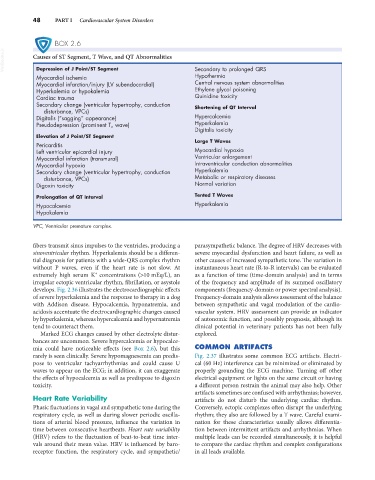
BOX 2.6
VetBooks.ir Causes of ST Segment, T Wave, and QT Abnormalities Secondary to prolonged QRS
Depression of J Point/ST Segment
Myocardial ischemia Hypothermia
Myocardial infarction/injury (LV subendocardial) Central nervous system abnormalities
Hyperkalemia or hypokalemia Ethylene glycol poisoning
Cardiac trauma Quinidine toxicity
Secondary change (ventricular hypertrophy, conduction Shortening of QT Interval
disturbance, VPCs)
Digitalis (“sagging” appearance) Hypercalcemia
Pseudodepression (prominent T a wave) Hyperkalemia
Digitalis toxicity
Elevation of J Point/ST Segment
Large T Waves
Pericarditis
Left ventricular epicardial injury Myocardial hypoxia
Myocardial infarction (transmural) Ventricular enlargement
Myocardial hypoxia Intraventricular conduction abnormalities
Secondary change (ventricular hypertrophy, conduction Hyperkalemia
disturbance, VPCs) Metabolic or respiratory diseases
Digoxin toxicity Normal variation
Prolongation of QT Interval Tented T Waves
Hypocalcemia Hyperkalemia
Hypokalemia
VPC, Ventricular premature complex.
fibers transmit sinus impulses to the ventricles, producing a parasympathetic balance. The degree of HRV decreases with
sinoventricular rhythm. Hyperkalemia should be a differen- severe myocardial dysfunction and heart failure, as well as
tial diagnosis for patients with a wide-QRS complex rhythm other causes of increased sympathetic tone. The variation in
without P waves, even if the heart rate is not slow. At instantaneous heart rate (R-to-R intervals) can be evaluated
+
extremely high serum K concentrations (>10 mEq/L), an as a function of time (time-domain analysis) and in terms
irregular ectopic ventricular rhythm, fibrillation, or asystole of the frequency and amplitude of its summed oscillatory
develops. Fig. 2.36 illustrates the electrocardiographic effects components (frequency-domain or power spectral analysis).
of severe hyperkalemia and the response to therapy in a dog Frequency-domain analysis allows assessment of the balance
with Addison disease. Hypocalcemia, hyponatremia, and between sympathetic and vagal modulation of the cardio-
acidosis accentuate the electrocardiographic changes caused vascular system. HRV assessment can provide an indicator
by hyperkalemia, whereas hypercalcemia and hypernatremia of autonomic function, and possibly prognosis, although its
tend to counteract them. clinical potential in veterinary patients has not been fully
Marked ECG changes caused by other electrolyte distur- explored.
bances are uncommon. Severe hypercalcemia or hypocalce-
mia could have noticeable effects (see Box 2.6), but this COMMON ARTIFACTS
rarely is seen clinically. Severe hypomagnesemia can predis- Fig. 2.37 illustrates some common ECG artifacts. Electri-
pose to ventricular tachyarrhythmias and could cause U cal (60 Hz) interference can be minimized or eliminated by
waves to appear on the ECG; in addition, it can exaggerate properly grounding the ECG machine. Turning off other
the effects of hypocalcemia as well as predispose to digoxin electrical equipment or lights on the same circuit or having
toxicity. a different person restrain the animal may also help. Other
artifacts sometimes are confused with arrhythmias; however,
Heart Rate Variability artifacts do not disturb the underlying cardiac rhythm.
Phasic fluctuations in vagal and sympathetic tone during the Conversely, ectopic complexes often disrupt the underlying
respiratory cycle, as well as during slower periodic oscilla- rhythm; they also are followed by a T wave. Careful exami-
tions of arterial blood pressure, influence the variation in nation for these characteristics usually allows differentia-
time between consecutive heartbeats. Heart rate variability tion between intermittent artifacts and arrhythmias. When
(HRV) refers to the fluctuation of beat-to-beat time inter- multiple leads can be recorded simultaneously, it is helpful
vals around their mean value. HRV is influenced by baro- to compare the cardiac rhythm and complex configurations
receptor function, the respiratory cycle, and sympathetic/ in all leads available.