
Page 1258 - Veterinary Immunology, 10th Edition
P. 1258
and TLR8 are more active in females than males. The immune
VetBooks.ir responses in lupus are closely associated with IFN-α production,
and the level of this cytokine correlates with disease activity. IFN-α
also promotes inflammation by activating macrophages and
autoreactive T cells, and many of the immunologic or pathogenic
features of lupus are driven by IFN-α. The production of ANAs in
lupus may also result if TLR7 and TLR9 lose the ability to
discriminate between microbial and self-DNA. If, at the same time,
some of their B cells undergo somatic mutation that enables their
BCRs to bind self-DNA, the ingredients necessary for a profound
antibody response to mammalian DNA come together.
Antinuclear antibodies can bind soluble nuclear antigens to form
immune complexes that are deposited in glomeruli, resulting in
development of a membranoproliferative glomerulonephritis
(MPGN) (Chapter 32). These immune complexes can also activate
neutrophils, causing them to release even more DNA and
nucleoproteins through NETosis. The immune complexes may also
be deposited in arteriolar walls, where they cause fibrinoid necrosis
and fibrosis, or in synovia, where they provoke arthritis.
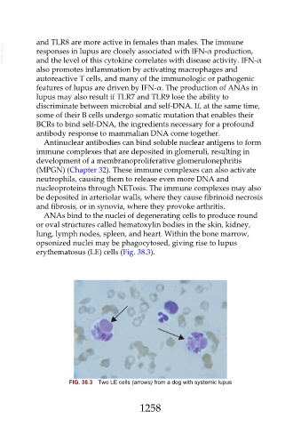
ANAs bind to the nuclei of degenerating cells to produce round
or oval structures called hematoxylin bodies in the skin, kidney,
lung, lymph nodes, spleen, and heart. Within the bone marrow,
opsonized nuclei may be phagocytosed, giving rise to lupus
erythematosus (LE) cells (Fig. 38.3).
FIG. 38.3 Two LE cells (arrows) from a dog with systemic lupus
1258