
Page 1411 - Veterinary Immunology, 10th Edition
P. 1411
VetBooks.ir
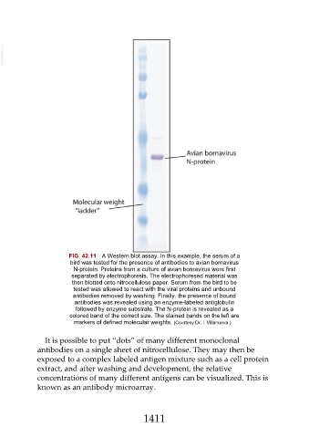
FIG. 42.11 A Western blot assay. In this example, the serum of a
bird was tested for the presence of antibodies to avian bornavirus
N-protein. Proteins from a culture of avian bornavirus were first
separated by electrophoresis. The electrophoresed material was
then blotted onto nitrocellulose paper. Serum from the bird to be
tested was allowed to react with the viral proteins and unbound
antibodies removed by washing. Finally, the presence of bound
antibodies was revealed using an enzyme-labeled antiglobulin
followed by enzyme substrate. The N-protein is revealed as a
colored band of the correct size. The stained bands on the left are
markers of defined molecular weights. (Courtesy Dr. I. Villanueva.)
It is possible to put “dots” of many different monoclonal
antibodies on a single sheet of nitrocellulose. They may then be
exposed to a complex labeled antigen mixture such as a cell protein
extract, and after washing and development, the relative
concentrations of many different antigens can be visualized. This is
known as an antibody microarray.
1411