
Page 1413 - Veterinary Immunology, 10th Edition
P. 1413
can be stained so that structural relationships are easier to see.
VetBooks.ir
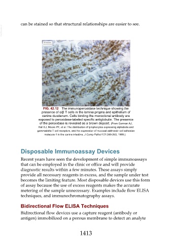
FIG. 42.12 The immunoperoxidase technique showing the
presence of α/β T cells in the lamina propria and epithelium of
canine duodenum. Cells binding the monoclonal antibody are
exposed to peroxidase-labeled specific antiglobulin. The presence
of the peroxidase is revealed as a brown deposit. (From German AJ,
Hall EJ, Moore PF, et al: The distribution of lymphocytes expressing alphabeta and
gammadelta T cell receptors, and the expression of mucosal addressin cell adhesion
molecule-1 in the canine intestine, J Comp Pathol 121:249-263, 1999.)
Disposable Immunoassay Devices
Recent years have seen the development of simple immunoassays
that can be employed in the clinic or office and will provide
diagnostic results within a few minutes. These assays simply
provide all necessary reagents in excess, and the sample under test
becomes the limiting feature. Most disposable devices use this form
of assay because the use of excess reagents makes the accurate
metering of the sample unnecessary. Examples include flow ELISA
techniques, and immunochromatography assays.
Bidirectional Flow ELISA Techniques
Bidirectional flow devices use a capture reagent (antibody or
antigen) immobilized on a porous membrane to detect an analyte
1413