
Page 1416 - Veterinary Immunology, 10th Edition
P. 1416
minutes.
VetBooks.ir
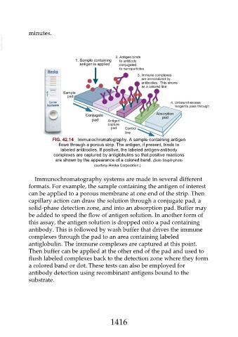
FIG. 42.14 Immunochromatography. A sample containing antigen
flows through a porous strip. The antigen, if present, binds to
labeled antibodies. If positive, the labeled antigen-antibody
complexes are captured by antiglobulins so that positive reactions
are shown by the appearance of a colored band. (Solo Step® photo
courtesy Heska Corporation.)
Immunochromatography systems are made in several different
formats. For example, the sample containing the antigen of interest
can be applied to a porous membrane at one end of the strip. Then
capillary action can draw the solution through a conjugate pad, a
solid-phase detection zone, and into an absorption pad. Buffer may
be added to speed the flow of antigen solution. In another form of
this assay, the antigen solution is dropped onto a pad containing
antibody. This is followed by wash buffer that drives the immune
complexes through the pad to an area containing labeled
antiglobulin. The immune complexes are captured at this point.
Then buffer can be applied at the other end of the pad and used to
flush labeled complexes back to the detection zone where they form
a colored band or dot. These tests can also be employed for
antibody detection using recombinant antigens bound to the
substrate.
1416