
Page 280 - The Veterinary Laboratory and Field Manual 3rd Edition
P. 280
Microbiology 249
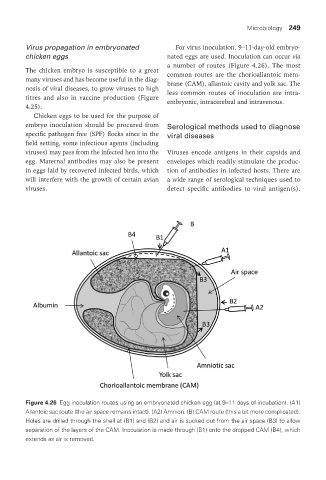
Virus propagation in embryonated For virus inoculation, 9–11-day-old embryo-
chicken eggs nated eggs are used. Inoculation can occur via
a number of routes (Figure 4.26). The most
The chicken embryo is susceptible to a great common routes are the chorioallantoic mem-
many viruses and has become useful in the diag- brane (CAM), allantoic cavity and yolk sac. The
nosis of viral diseases, to grow viruses to high less common routes of inoculation are intra-
titres and also in vaccine production (Figure embryonic, intracerebral and intravenous.
4.25).
Chicken eggs to be used for the purpose of
embryo inoculation should be procured from Serological methods used to diagnose
specific pathogen free (SPF) flocks since in the viral diseases
field setting, some infectious agents (including
viruses) may pass from the infected hen into the Viruses encode antigens in their capsids and
egg. Maternal antibodies may also be present envelopes which readily stimulate the produc-
in eggs laid by recovered infected birds, which tion of antibodies in infected hosts. There are
will interfere with the growth of certain avian a wide range of serological techniques used to
viruses. detect specific antibodies to viral antigen(s).
Figure 4.26 Egg inoculation routes using an embryonated chicken egg (at 9–11 days of incubation). (A1)
Allantoic sac route (the air space remains intact). (A2) Amnion. (B) CAM route (this a bit more complicated).
Holes are drilled through the shell at (B1) and (B2) and air is sucked out from the air space (B3) to allow
separation of the layers of the CAM. Inoculation is made through (B1) onto the dropped CAM (B4), which
extends as air is removed.
Vet Lab.indb 249 26/03/2019 10:25